Jefferson Fracture
Brett R. Murdock
Daniel B. Nissman
CLINICAL HISTORY
24-year-old male presents with pain in the upper neck and limited range of motion after cliff diving in shallower than expected water.
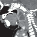
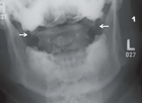 FIGURE 60A |
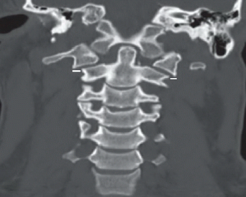 FIGURE 60B |
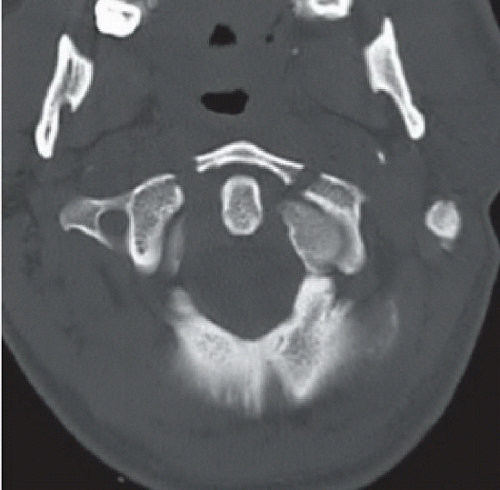 FIGURE 60C |
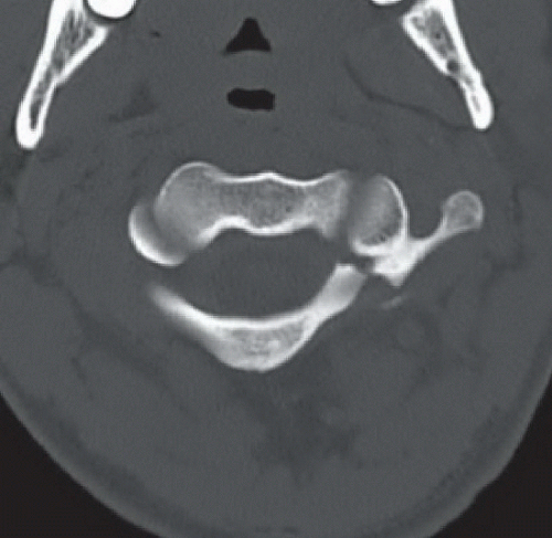 FIGURE 60D |
FINDINGS
Anteroposterior (AP) radiograph (Fig. 60A) demonstrates lateral offset of the lateral masses of C1 relative to the lateral margins of C2 (arrows). Coronal reconstruction from cervical spine CT (Fig. 60B) confirming lateralization of the lateral masses of C2 relative to the occipital condyles as well as the lateral margins of C2. Short white lines indicate the degree of lateralization of the lateral masses of C1; if the sum is greater than or equal to 7 mm, transverse ligament disruption is likely. Axial images from cervical spine CT confirm two anterior C1 ring fractures (Fig. 60C) and a displaced fracture of the left lateral C1 ring (Fig. 60D). (Images courtesy of Jordan Renner, MD, University of North Carolina, Chapel Hill, NC, USA.)
DIFFERENTIAL DIAGNOSIS
Congenital variant (cleft/malformation), C1 ring fracture (Jefferson).
Related posts:


Stay updated, free articles. Join our Telegram channel

Full access? Get Clinical Tree


