Knee radiographs are widely used in clinical practice. Many features can be depicted when a systematic analysis of the different views is performed. This article focuses on different types of joint effusion and on the analysis of the bone outlines of the knee, particularly on the lateral view. Systematic analysis of these bone outlines and knowledge of several key points are particularly useful for the depiction of abnormal bone morphology or positioning, and of several conditions, such as trochlear dysplasia, patellar dislocation, impaction fractures, or ligament injuries and avulsion fractures.
Key points
- •
Joint effusion, lipohemarthrosis, and exuberant subsynovial frondlike fatty proliferation in lipoma arborescens can be depicted on a lateral view.
- •
Normal femoral notches should be differentiated from the lateral femoral notch sign associated with anterior cruciate ligament tears.
- •
Trochlear dysplasia and features reflecting its severity should be systematically sought after.
- •
Patella alta, patellar tilt, and complete lateral patellar dislocation can be associated with trochlear dysplasia.
- •
Analysis of the bone outlines can allow depiction of small avulsion fractures.
Knee radiographs are still widely used in clinical practice. Many features can be depicted when a systematic analysis of the different views is performed. In this article, after discussing different types of joint effusion, we focus on the analysis of the bone outlines of the knee, particularly on the lateral view. Systematic analysis of these bone outlines and knowledge of several key points are particularly useful for the depiction of abnormal bone morphology or positioning, and of several injuries.
Joint effusion
Whatever the clinical context, the presence of a joint effusion should be systematically looked for on a lateral knee radiograph. This increased intra-articular amount of fluid is not specific of a particular disorder, as can occur in a variety of settings. However, it draws attention to the presence of a joint disorder. It leads to the separation between the suprapatellar and prefemoral fat pads by a well-defined rounded suprapatellar recess ( Fig. 1 ). Loss of normal posterior fat plane of the quadriceps tendon and anterior displacement of the latter and of the patella are only seen in large joint effusions.

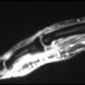
In trauma, a fat-fluid level related to lipohemarthrosis can be seen when a lateral knee view with horizontal ray is performed ( Fig. 2 ) or on a lateral knee view performed in a standing patient ; it is specific for the existence of an intra-articular fracture, allowing bone marrow fat to leak into the joint via the fracture and to float on top of the blood. The detection of this sign is particularly useful when the intra-articular extent of a fracture is not obvious on plain films, as it should lead to additional imaging assessment.

Sometimes, fatty lucencies that seem to be entrapped in a distended suprapatellar pouch can be detected on radiographs ( Fig. 3 ). They are related to extensive subsynovial frondlike fatty proliferation in lipoma arborescens and can be depicted radiographically when exuberant. This latter disorder is typically associated with chronic arthropathies, such as osteoarthritis or rheumatoid arthritis.

Lateral femoral notch sign
Normal Anatomy
On a true lateral view, the 2 femoral condyles are superimposed, particularly their posterior border. However, analysis of their shape allows differentiating them: the middle third of the lateral condyle is flat and may demonstrate a mild notch, whereas the medial condyle is more rounded. When the superposition of the 2 condyles is not perfect, which is frequently the case, a large notch also can be recognized at the junction between the anterior and middle third of the medial condyle. These normal notches, also known as condylopatellar sulcus, represent the junction zone on the femoral condyles where the tibiofemoral and patellofemoral radii of curvature meet ( Fig. 4 ).

Lateral Femoral Notch Sign
Normal notches should be differentiated from the lateral femoral notch sign, which results from the impaction between the lateral femoral condyle and the posterior aspect of the tibial plateau during anterior cruciate ligament tears ( Fig. 5 ). This inconstant osteochondral fracture involves the middle to anterior portion of the lateral femoral condyle; it can be recognized radiographically when it is deeper (>1.5 mm) and/or has sharper angulation than the normal lateral notch (see Fig. 5 ).

Trochlear dysplasia
This developmental anomaly is one of the most important risk factors for lateral patellar dislocation. Its depiction relies on the analysis of the proximal part of the trochlea on a true lateral radiograph, as it has been demonstrated that this view is more reliable for the diagnosis of trochlear dysplasia than axial radiographs obtained at 30° of flexion of the knee. Thus, it is crucial for diagnostic accuracy of trochlear dysplasia to have a correct lateral radiograph with superimposition of the femoral condyles posteriorly, as a rotational deviation of at least 5° can cause false-positive or false-negative diagnosis of trochlear dysplasia.
Normal Anatomy
Normally, the ventral outline of both femoral condyles projects at a distance from the tangent to the deepest part of the trochlear groove and should not cross it, as both condyles continue with the ventral cortical bone of the femoral metaphysis. This means there is a normal shape and depth of the proximal trochlea ( Fig. 6 ). The depth of the proximal trochlea can be assessed 1 cm below the proximal end of the trochlear groove and should be more than 5 mm (see Fig. 6 ). This measure is rarely performed in clinical practice, but it constitutes a good landmark for normal anatomy in cases of doubt.

Trochlear Dysplasia
Trochlear dysplasia is defined by the presence of the crossing sign, when the tangent to the deepest part of the trochlear groove crosses the ventral outline of the lateral condyle ( Fig. 7 ). At this level, the trochlea becomes flat, which may lead to patellar instability, as the patella cannot engage normally in the trochlea.

It is important to keep in mind that in a normal knee, the lateral femoral condyle extends higher anteriorly than the medial condyle. This feature can be used to differentiate the 2 condyles, but it also explains that, quite frequently, the ventral outline of the medial condyle joins the tangent to the trochlear groove, as the proximal part of the medial trochlear facet can be in a frontal plane. This is a normal and frequent feature; this is not a crossing sign ( Fig. 8 ). The crossing sign must involve at least the lateral condyle. This feature should be kept in mind to avoid overdiagnosis of trochlear dysplasia.

Features Reflecting the Severity of the Trochlear Dysplasia
Once the crossing sign has been recognized, other features reflecting the severity of the trochlear dysplasia should be systematically sought out :
- •
A supratrochlear spur or bump , which plays a role similar to a ski jump when the patella engages in the trochlea ( Fig. 9 ). This feature usually indicates ventral prominence of the trochlea, which can be measured by the distance between a line drawn through the ventral cortical bone of the femoral shaft and a parallel line tangent to the most ventral point of the trochlear floor. This measure is approximately 0 to 2 mm in normal knees but can be more than 3 mm in severe trochlear dysplasia ( Fig. 10 ). The trochlea is usually markedly flattened or even convex in this region.