Joint Injection: Upper Limb
KEY FACTS
Procedure
 Radiocarpal, distal radioulnar, & midcarpal joints
 Identify & avoid both superficial branch of radial nerve & terminal part posterior interosseous nerve

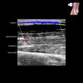
The needle tip should just pass through the capsule and synovium into the superficial aspect of the joint. The synovium can be difficult to see clearly if the joint is not distended by fluid. Place the needle tip in the joint cavity between the opposing articular cartilage surfaces.

The needle tip should not be forced deeply into the joint, particularly if the tip is not well seen as this may cause the injury to the articular cartilage, as shown.

One can place the needle tip onto the “‘bare area”  covered by synovium, although it is devoid of articular cartilage (blue needle) as injection can be closely monitored. Not every joint has a “bare area” and alternatively, one can place the needle tip just superficial to the articular cartilage (yellow needle).

For an out-of-plane approach, to avoid partial leakage of injectant outside the joint (upper diagram), one can use either a needle with a short bevel (left lower) or align the needle obliquely (right lower).
PROCEDURE
Procedure Steps (General)
 Ideally, needle tip should just pass through synovium & be within superficial part of joint
 Synovium is difficult to separate from subsynovial fat if there is no joint effusion
 Contacting cartilage with needle tip is not harmful
 One can often locate “bare area” between capsule & cartilage where needle tip can be positioned
 If moderate to large effusion present, aspirate joint fluid prior to injection
Shoulder (Glenohumeral Joint)
 Less painful, more comfortable position to patient than posterior approach
 Patient in supine position with arm partially externally rotated
 Biceps tendon runs in rotator interval between subscapularis & supraspinatus tendons
 Identify coracoid process, which is medial to rotator interval
 Needle entry point is just superior to subscapularis tendon, inferior to supraspinatus tendon, & lateral to coracoid process
 Needle position should be medial to biceps tendon
 Needle is advanced until tip just superficial to articular cartilage of humeral head
 Either short-axis or long-axis needle approach can be used
Elbow Joint
 More conventional, classic method & ergonomic in terms of patient positioning, but more difficult & time-consuming to locate injection site
 Patient sits next to table with flexed elbow resting on table & lateral aspect uppermost
 Position probe longitudinally along lateral part of elbow
![]()
Stay updated, free articles. Join our Telegram channel

Full access? Get Clinical Tree