, Maria Custódia Machado Ribeiro2 and Bruno Beber Machado3
(1)
Hospital da Criança de Brasília – Jose Alencar, Clínica Vila Rica, Brazília, Brazil
(2)
Hospital da Criança de Brasília – Jose Alencar, Hospital de Base do Distrito Federal, Brasília, Brazil
(3)
Clínica Radiológica Med Imagem, Unimed Sul Capixaba, Santa Casa de Misericordia de, Cachoeiro de Itapemirim, Brazil
Abstract
The term juvenile idiopathic arthritis (JIA) encompasses a heterogeneous group of chronic arthritides that share the following features: (1) disease onset before 16 years of age and (2) a minimum duration of 6 weeks. For many years, it was named juvenile rheumatoid arthritis (mainly in the American literature) and chronic juvenile arthritis (mostly in European texts). It is an autoimmune inflammatory disease of unknown etiology that occurs most often in females, whose main target tissue is the synovium. Synovitis is present since the very early stages of JIA, with gradual development of synovial hyperplasia and hypercellular pannus, which eventually erodes cartilage and bone. Even though this diagnosis is essentially a clinical one, recent therapeutic advances have drastically changed the role of imaging in its management. Children with JIA must be diagnosed as soon as possible, aiming at a benign course and a better outcome with timely introduction of treatment. Magnetic resonance imaging is especially useful as it allows for early demonstration of abnormal findings, monitoring of disease progression, and accurate assessment of treatment response.
3.1 Introduction
The term juvenile idiopathic arthritis (JIA) encompasses a heterogeneous group of chronic arthritides that share the following features: (1) disease onset before 16 years of age and (2) a minimum duration of 6 weeks. For many years, it was named juvenile rheumatoid arthritis (mainly in the American literature) and chronic juvenile arthritis (mostly in European texts). It is an autoimmune inflammatory disease of unknown etiology that occurs most often in females, whose main target tissue is the synovium. Synovitis is present since the very early stages of JIA, with gradual development of synovial hyperplasia and hypercellular pannus, which eventually erodes cartilage and bone. Even though this diagnosis is essentially a clinical one, recent therapeutic advances have drastically changed the role of imaging in its management. Children with JIA must be diagnosed as soon as possible, aiming at a benign course and a better outcome with timely introduction of treatment. Magnetic resonance imaging is especially useful as it allows for early demonstration of abnormal findings, monitoring of disease progression, and accurate assessment of treatment response.
The classification of JIA is a complex and somewhat controversial issue, and a thorough analysis of it is beyond the scope of this chapter. The following topics will cover the imaging findings of peripheral joint disease in the classic form of JIA. The juvenile spondyloarthropathies will be the discussed in Chap. 4, while spinal involvement in JIA is part of the subject matter of Chap. 11.
3.2 Radiographs
For many decades, radiographs were the only imaging modality available for the assessment of patients with JIA. The accumulated experience with this method is quite vast, so that radiographic findings in advanced disease are characteristic. Nevertheless, radiographs are fairly insensitive for the early stages of arthritides in general, underestimating the real severity of joint damage. This is particularly true for JIA because the epiphyseal cartilage of the long bones is thick and extensive cartilaginous loss must occur before bone erosions become apparent in children. Despite these limitations, radiographs must be requested for all patients as part of the initial assessment (baseline study), being also useful to rule out alternative causes of joint pain.
Early-stage changes, such as synovial thickening and joint effusion, appear on radiographs as nonspecific soft-tissue swelling and obliteration of fat planes (Fig. 3.1). Periarticular soft-tissue swelling is more evident if the joint is surrounded by air and a natural density gradient is therefore present (Fig. 3.2). Findings related to chronic inflammation and hyperemia include juxta-articular osteoporosis (Fig. 3.3), epiphyseal remodeling, and increased size of secondary ossification centers (Fig. 3.4). The hyperemic state may also lead to accelerated skeletal maturation and divergence between bone age – as assessed by radiographs – and the actual chronological age. Bone overgrowth and/or early closure of the growth plates (Figs. 3.4, 3.5, 3.6, and 3.7) can be found, as well as limb-length discrepancy (Fig. 3.8).
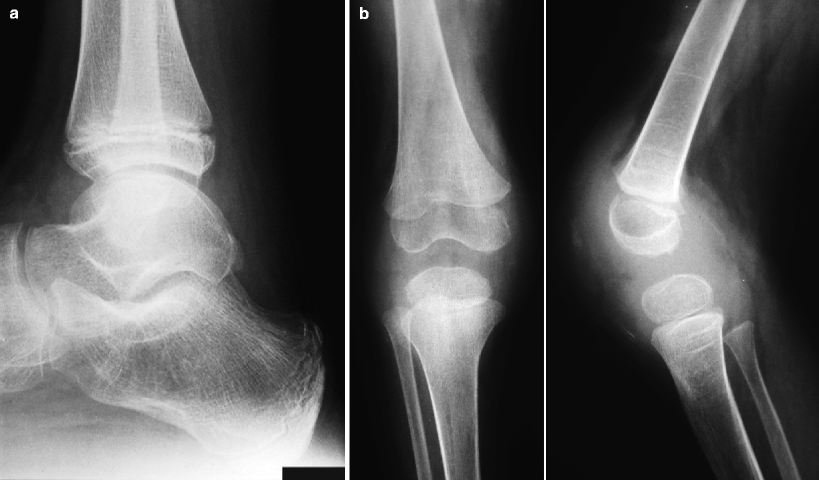
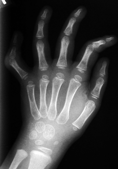
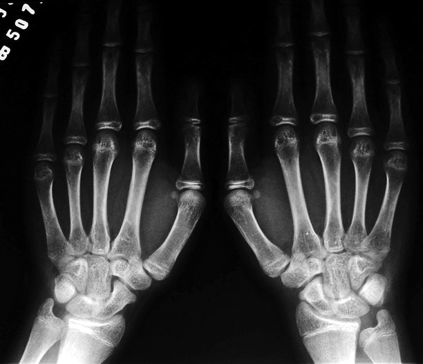
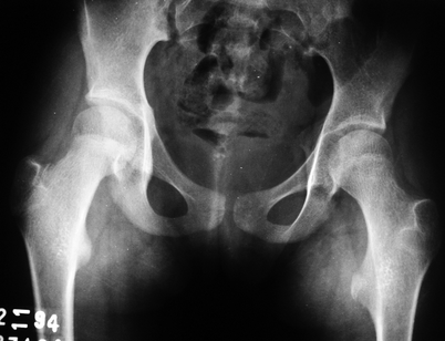
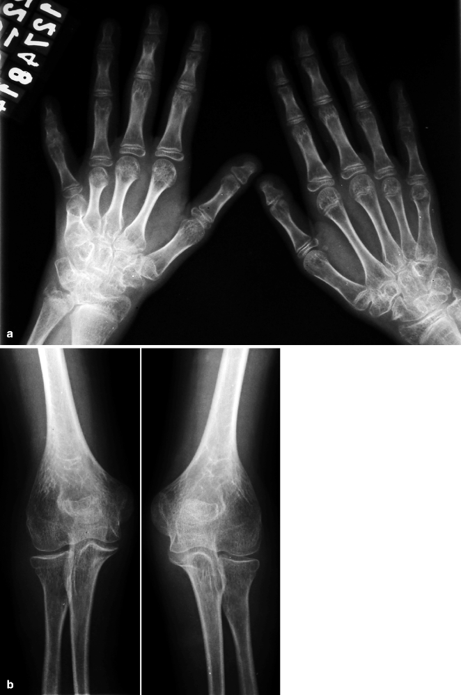
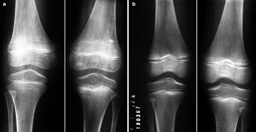
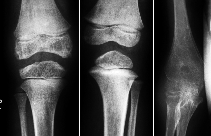
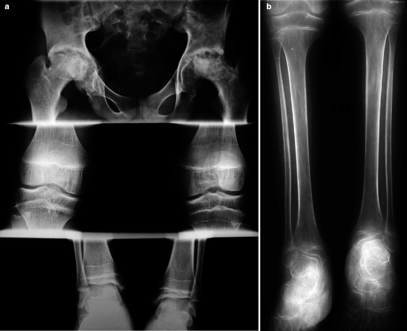

Fig. 3.1
Radiographs of the ankle (a) and of the knee (b) of two children with JIA showing soft-tissue swelling and displacement of periarticular fat planes. Even though bone erosions are not apparent, osteoporosis is already present, notably in the knee, where soft-tissue swelling is much more prominent

Fig. 3.2
Radiograph of the right hand of a child with JIA. There is soft-tissue swelling adjacent to the proximal interphalangeal joint of the fifth finger, more evident due to the density gradient created by the air/fat interface. Periarticular osteoporosis and periosteal reaction along the diaphyses of the proximal phalanges are also present

Fig. 3.3
Radiographs of the hands and wrists of a child with early-stage JIA. Symmetric and bilateral periarticular osteoporosis is the main finding, predominantly found in the metacarpophalangeal joints. There is also subtle increase in the size of the epiphyses, mainly in the left wrist, without signs of erosive articular disease

Fig. 3.4
Radiograph of the pelvis of a patient with long-standing JIA displaying findings related to chronic hyperemia. The proximal epiphysis of the right femur shows increased size, while there is diffuse osteoporosis associated with hypoplasia of the bones and early physeal closure at left. Bilateral coxa valga is also present

Fig. 3.5
Radiograph of the hands of a patient with long-standing JIA (a) discloses acquired bilateral deformity of several digits due to premature and asymmetric closure of the growth plates, with shortening of the second to the fifth metacarpals at right and of the distal phalanges of both thumbs. Diffuse osteoporosis is also present, with abnormal alignment of the carpal bones and increased size of several epiphyses, bilaterally. Narrowing of joint spaces is obvious, mainly in the right radiocarpal joint, with invagination of the adjacent carpal bones. In (b), in another child with late-stage JIA, there is growth arrest due to premature closure of most physes in the elbows. Marked periarticular osteoporosis is also present, with increased size of the distal humeral epiphyses, notably in the left elbow

Fig. 3.6
In (a), anteroposterior view shows asymmetric growth of the knees, with increased size of the left distal femur when compared to the contralateral (which also displays mild overgrowth); diffuse osteoporosis and growth recovery lines can be seen, bilaterally. In (b), only the left knee is affected, with epiphyseal overgrowth of the distal femur and of the proximal tibia and mild homolateral osteoporosis; growth recovery lines are also present

Fig. 3.7
The left image reveals asymmetric bone maturation in the knees of a child with JIA: the epiphyses of the right femur, tibia, and fibula are irregular, bigger, and more developed than the contralateral, and regional osteoporosis is also present. In the right image, there is diffuse osteoporosis of the elbow, marked periosteal reaction along the distal humerus, and epiphyseal overgrowth. Erosive articular disease is evident, mainly in the medial portion of the joint, as well as soft-tissue swelling

Fig. 3.8
Scanogram of an adolescent with advanced JIA (a) demonstrating advanced hip arthritis, bilaterally, more important at left, with widespread erosions and remodeling of the joint surfaces. There is shortening of the right limb, with marked leg-length discrepancy. Growth recovery lines and periarticular osteoporosis can be seen in the knees and ankles. Anteroposterior view of the lower limbs of another patient with JIA (b) shows diffuse osteoporosis and increased size of the epiphyses of the knees and ankles; the left limb is shorter than the contralateral as a result of abnormalities above the knee level. Tibiotalar slant can be seen, bilaterally
The so-called joint space seen on radiographs comprises, in fact, the joint cavity itself and non-calcified structures (which are cartilaginous for the most part) interposed between the joint surfaces. This space undergoes slow, progressive, and uniform narrowing due to the action of pannus, which erodes and destroys the cartilaginous structures (Figs. 3.9 and 3.10). Bone erosions are first seen in peripheral sites where the articular cartilage is absent, near to ligament insertions and capsular reflections (Fig. 3.11), presenting centripetal progression. Erosions in the joint surfaces are indicative of advanced disease, which may lead to secondary osteoarthritis (Figs. 3.8, 3.12, and 3.13). Destruction of the joint surfaces may eventually lead to ankylosis, infrequently found nowadays (Fig. 3.14). Periosteal reaction is more frequently encountered in JIA than in the adult form of rheumatoid arthritis, affecting mostly the tubular bones of hands and feet (Figs. 3.2, 3.7, 3.10, 3.14, and 3.15). Bone spurs and enthesophytes are associated with enthesitis (inflammation involving the sites of insertion of tendons, ligaments, and joint capsules).
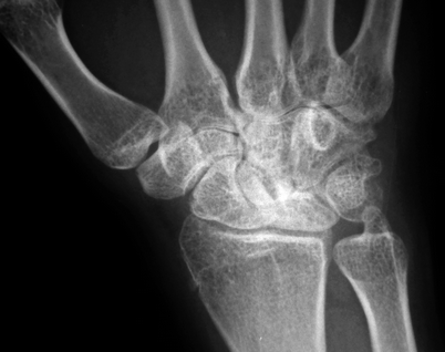
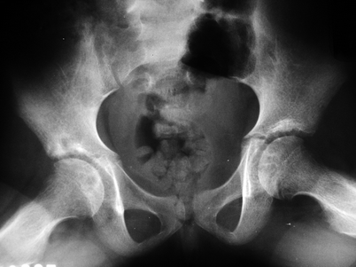
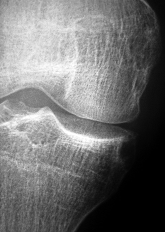
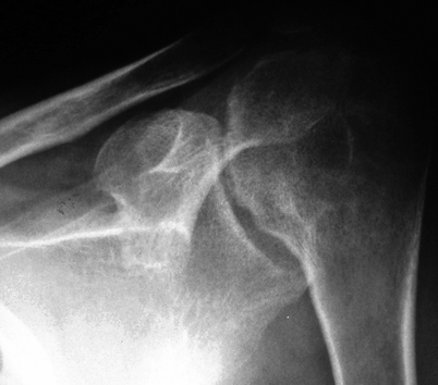
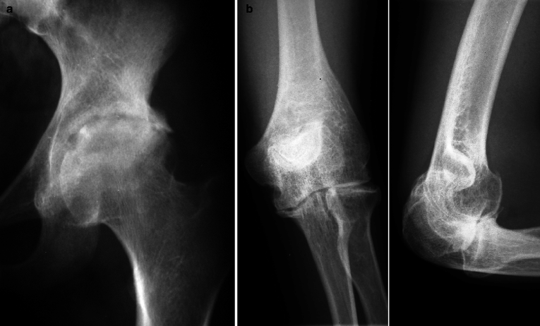
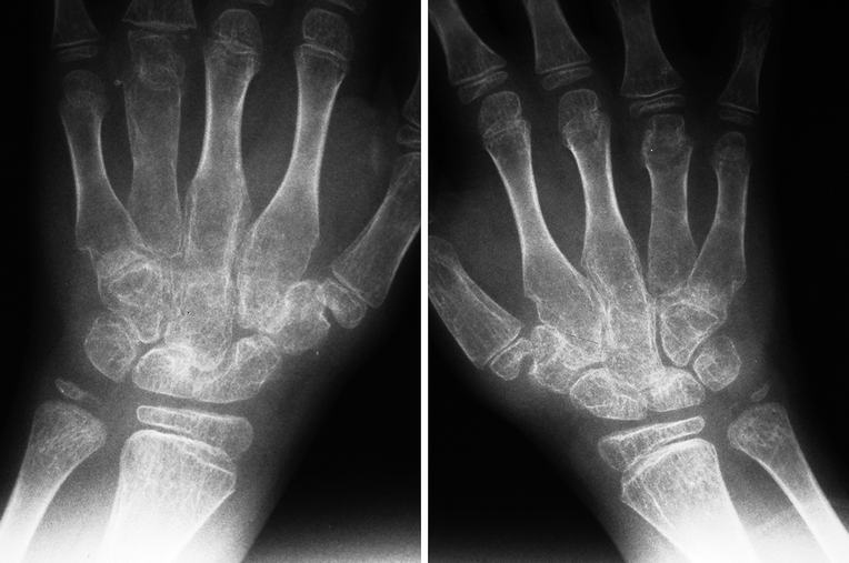
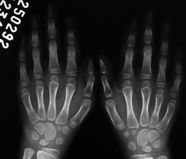

Fig. 3.9
Radiograph of the left wrist of a 16-year-old female with JIA since late childhood. There is mild osteoporosis, with narrowing of the radiocarpal joint space and disorganization of the proximal carpal row; a small cortical erosion can be seen in the distal third of the scaphoid. Despite the absence of extensive bone erosions, these findings are indicative of advanced cartilaginous destruction

Fig. 3.10
Long-standing JIA affecting both hips, with bone erosions, narrowing of the joint spaces (more important in the right hip), and periarticular osteoporosis. Periosteal reaction is seen along the right femoral neck

Fig. 3.11
Radiograph of the right knee of an adolescent with JIA and erosive changes. A large peripheral bone erosion can be seen in the proximal tibia, not affecting the joint surface of the adjacent plateau

Fig. 3.12
Radiograph of the left shoulder of a patient with long-standing JIA. The joint surface of the humeral head is markedly irregular due to the presence of bone erosions. Cephalic migration of the humeral head is also present, related to complete tear of the rotator cuff. Glenoid deformity and bone remodeling are also evident

Fig. 3.13
In (a), radiograph of the left hip of a patient with advanced JIA demonstrates erosive arthropathy and secondary osteoarthritis due to extensive cartilaginous damage, with marked narrowing of the joint space associated with marginal osteophytes and subchondral sclerosis. In (b), radiographs of the left elbow of a 22-year-old patient, suffering from JIA since he was 1 year old, reveal premature osteoarthritis secondary to chronic arthritis

Fig. 3.14
Radiographs of the wrists of a 6-year-old child with JIA since she was 18 months old. There is diffuse osteoporosis and ankylosis of multiple joints, mainly carpometacarpal, as well as bilateral arthritis of the fourth metacarpophalangeal joints, abnormal bone modeling, and solid periosteal reaction in the metacarpal diaphyses

Fig. 3.15
Symmetric and bilateral periostitis can be seen along the proximal and middle phalanges in both hands, notably from the second to the fourth digits. Periarticular osteoporosis is also present in this patient with JIA
In advanced JIA, the association of diffuse osteoporosis (Fig. 3.16) and anomalous biomechanics (caused by changes such as muscle atrophy and abnormal joint alignment) increases the risk of fractures. Radiodense metaphyseal bands can be found in patients to whom bisphosphonates were administered to treat osteoporosis (Fig. 3.17). Growth recovery lines are frequently seen (Figs. 3.5, 3.6, 3.8, and 3.16), mostly in patients with long-standing disease, as well as the “bone-within-bone” appearance, which has a similar meaning (Fig. 3.18). Intra-articular and periarticular soft-tissue calcifications are also common, most often secondary to therapeutic corticosteroid injections (Fig. 3.19). Flexion contractures, varus/valgus deformities, and altered joint alignment are other late-stage complications (Fig. 3.20).
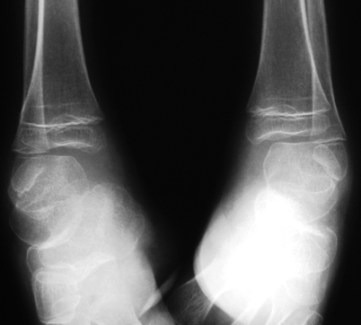
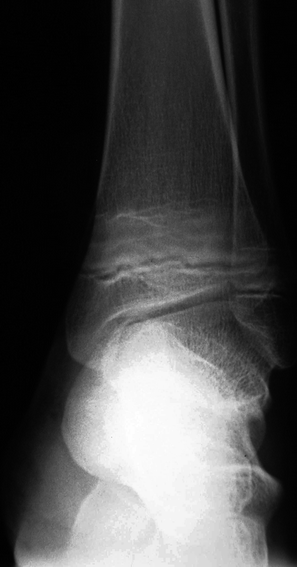

Fig. 3.16
There is marked widespread osteoporosis in both ankles in this patient with long-standing JIA, as well as soft-tissue swelling and growth recovery lines

Fig. 3.17
This 14-year-old patient developed diffuse osteoporosis after institution of corticotherapy for treatment of JIA, and treatment with oral alendronate was started. Radiographs taken 10 months later disclosed dense metaphyseal bands in his long bones, here shown in the left ankle. Tibiotalar slant is also evident

