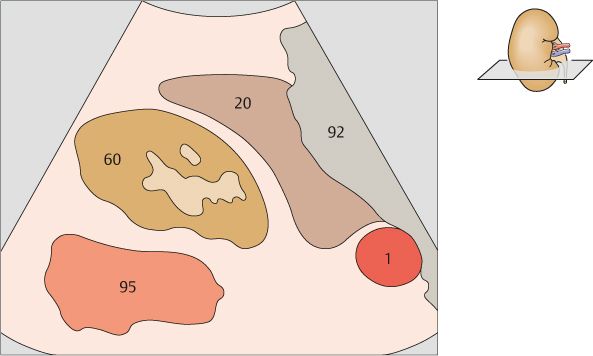
175 Kidney, liver, psoas muscle
176 Kidney, liver, psoas muscle

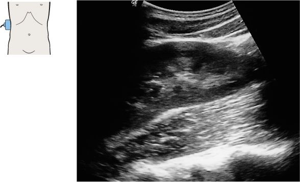
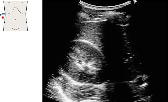
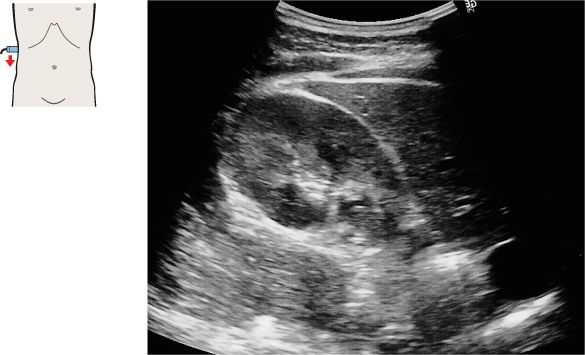
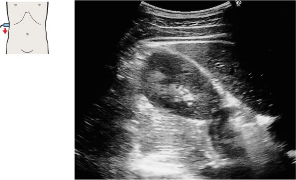
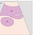
The right kidney is clearly demonstrated through the acoustic window of the liver.
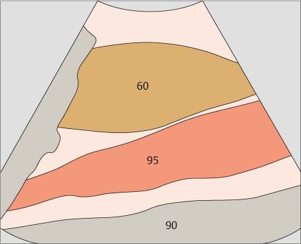
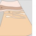
The kidneys slide downward along the lumbar muscles during respiratory excursions.
177 Kidney, liver, psoas muscle
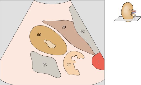
178 Kidney, right renal vein
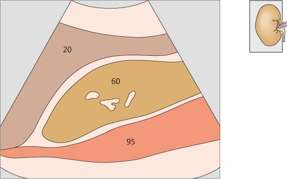
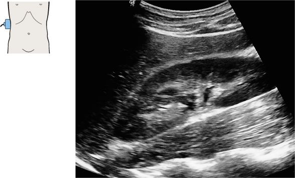
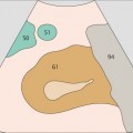
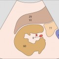
The summation of portions of the pelvicalyceal system, blood vessels, lymphatics, fatty tissue, and renal sinus form an echogenic complex at the center of the kidney.
The fibrous renal capsule cannot be visualized with ultrasound.
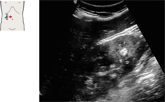
179 Kidney, liver
180 Kidney, liver, right renal vein
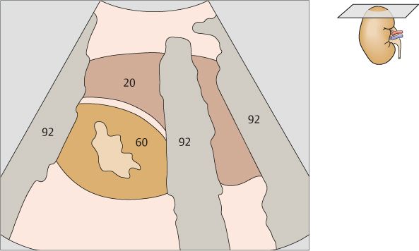
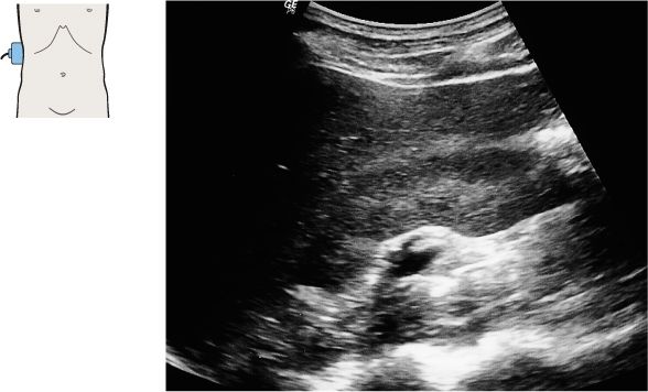
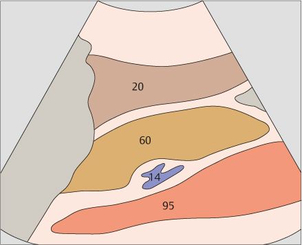
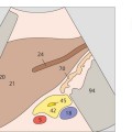
The right kidney occupies a posterior site in the angle between the spinal column, muscles, and right lobe of the liver.
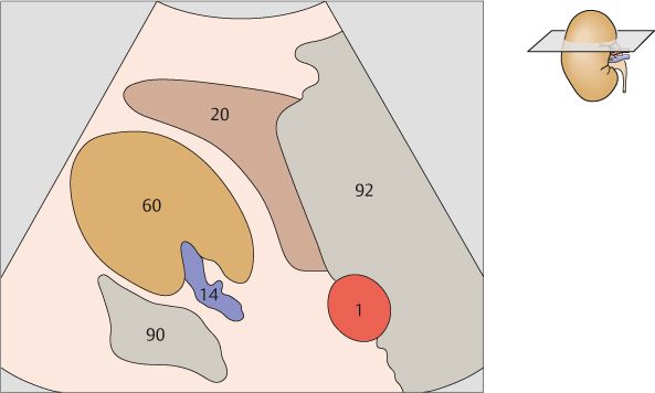
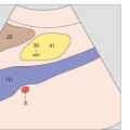
Generally the renal hilar vessels can be clearly defined.
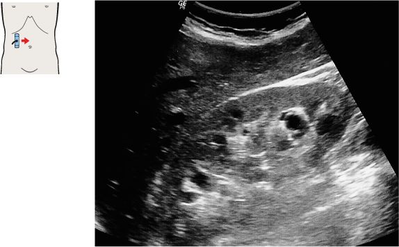
181 Kidney, liver
182 Kidney, liver
The psoas muscle is located medial to the kidney.
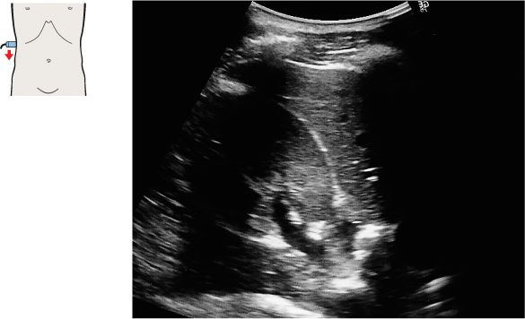
In most cases the right kidney can be clearly visualized as far as its upper pole when scanned from the lateral side.
183 Kidney, liver
184 Kidney, liver
Stay updated, free articles. Join our Telegram channel

Full access? Get Clinical Tree


