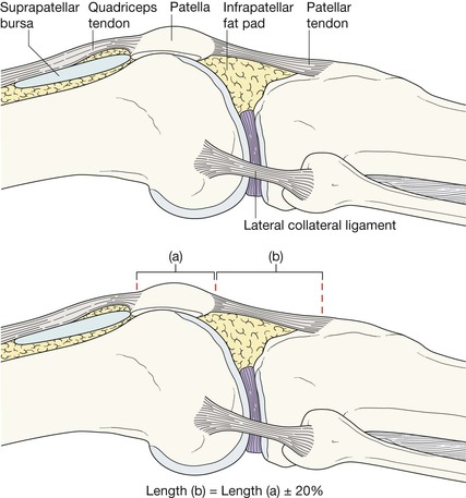
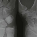
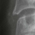
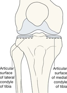
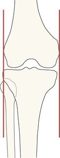
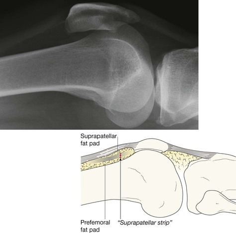
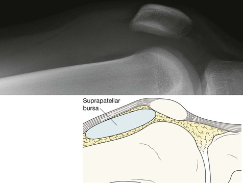
Check: 2. The head and neck of the fibula. □ Each plateau should be smooth. No steps, no layering, no disruptions. □ The subchondral bone should not show any focal increase in density. 4. The patella. Look through the superimposed femur. 5. Finally, check for any small fragments of bone—anywhere at all. Normal AP. Tibial articular surfaces. Appear as an oval on either side of the intercondylar eminence. These ovals should be smooth without any steps or layering. In the normal knee a perpendicular line drawn at the most lateral or medial margin of the femur should have no more than 5 mm of adjacent tibia outside of it. If this rule is broken suspect a plateau fracture. 1–5 as for adults, but also check: 6. The growth plates of femur, tibia, and fibula. Is there an epiphyseal fracture? (See pp. 14–17). 7. The cortex of the femur and tibia. Is there a Greenstick or Torus fracture? (See pp. 18–19.) 8. The condylar surfaces of the femur. Is there an osteochondral lesion/fracture? (See pp. 28–29.) Normal AP. It is important to understand the normal and abnormal appearance of the suprapatellar bursa—if abnormal, this often suggests a fracture or ligament damage4–6. Check: 1. For a joint effusion4–7. Present if the suprapatellar strip exceeds 5 mm (see below). 2. For a fat–fluid level in the suprapatellar bursa… an intra-articular fracture. 3. The condylar surfaces of the femur. Are they smooth? 4. The patella. Is the articular surface smooth? 5. The position of the patella. Large effusion. Note the marked differences compared with the normal appearances on the page opposite. The suprapatellar strip is markedly widened. Effusion with a fat–fluid level. In the Emergency Department the lateral view of the knee is obtained with a horizontal X-ray beam (conventionally referred to as a “HBL”). A fat–fluid level occurs when fat (arrowheads) lies on top of blood in the suprapatellar bursa. The fat has been released from bone marrow and consequently the fat–fluid level indicates an intra-articular fracture7–9.
Knee
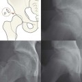
Normal anatomy
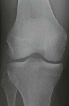
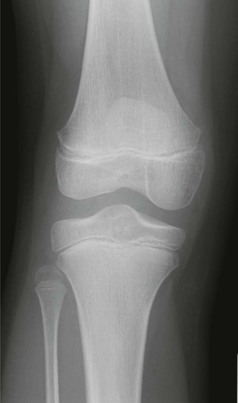
AP view
Analysis: the checklists
The AP radiograph
Adults: a five-point checklist
Children: an eight-point checklist
The lateral radiograph
Adults and children: a six-point checklist
Related posts:
![]()
Stay updated, free articles. Join our Telegram channel

Full access? Get Clinical Tree


Knee
15