and Clarisse Dromain2
(1)
Department of Radiology, San Giovanni Hospital, Roma, Italy
(2)
Department of Radiology, Institut de Cancerologie Gustav Roussy, VilleJuif – Paris, France
Abstract
The levator ani muscle is a broad thin muscle of the pelvis; it supports pelvic viscera and structures that pass it through. The levator ani muscle in conjunction with the coccygeal muscle forms the pelvic diaphragm.
Levator Ani Muscle
The levator ani muscle is a broad thin muscle of the pelvis; it supports pelvic viscera and structures that pass it through. The levator ani muscle in conjunction with the coccygeal muscle forms the pelvic diaphragm.
The levator ani muscle is divided into three parts: the ileococcygeal muscle, pubococcygeal, and puborectal muscle.
The contraction of all components of levator ani muscle realizes the rising up of the pelvic floor, and at the same time, thanks to the action of puborectal muscle, the anorectal junction is pushed anteriorly with reduction of anorectal angle.
The relaxation of levator ani muscle leads to the opening of anorectal angle. Therefore the action of levator ani muscle and the specific action of one of its branches (puborectal muscle) play an important role in defecation and fecal continence as well as in pelvic floor disease such as rectal prolapse, descending perineum syndrome, obstructed defecation syndrome, and abdominopelvic incoordination.
The levator ani muscle syndrome is characterized by episodical rectal pain (proctalgia fugax) due to spasm of the levator ani muscle.
The levator ani muscle is also an important anatomical landmark in categorization of perianal fistula particularly in the definition of suprasphincteric and extrasphincteric fistulas in which the levator ani muscle is involved in the fistulous tract.
MRI using phased-array coil, high magnetic field strength (1.5 T magnet) acquiring high spatial resolution TSE T2-weighted images on coronal and axial planes is the best imaging method to imagine the levator ani muscle from a morphologic point of view.
MR defecography is used to investigate functionality of the levator ani muscle as well as the defecation function.
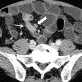
Lymphoma, GI Tract
Gastric and duodenal lymphomas are most often B-cell non-Hodgkin’s lymphoma (MNHL). Lymphomas may occur as a primary gastric lesion or as part of a disseminated disease. The primary location of MNHL in the stomach accounts for less than 5 % of malignant gastric tumors. However the stomach is the most common extranodal site of primary lymphomas.< div class='tao-gold-member'>Only gold members can continue reading. Log In or Register to continue

Stay updated, free articles. Join our Telegram channel

Full access? Get Clinical Tree


