Lacrimal system and salivary glands
Digital subtraction and CT dacryocystography
Dacryocystography allows visualization of the lacrimal system by direct injection of contrast into the canaliculus of the eyelid. It may be combined with CT imaging performed immediately following lacrimal system contrast injection. CT imaging gives additional diagnostic information, particularly about adjacent structures.1
Technique
1. The lacrimal sac is massaged to express its contents prior to injection of the contrast medium. The lower eyelid is everted to locate the lower canaliculus at the medial end of the lid.
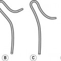
2. The lower punctum normally measures 0.3 mm in diameter and is gently dilated. The lower lid should be drawn laterally to straighten the curve in the canaliculus. To avoid perforation, the cannula is initially positioned perpendicular to the eyelid margin as it enters the punctum. The cannula is then rotated by 90° horizontally along the natural curve of the lower canaliculus.
3. Care is taken not to advance the cannula too far into the canaliculus as proximal stenosis can be missed.
4. The upper canaliculus may also be cannulated if there is difficulty with the lower canaliculus.
5. The contrast medium is injected and a digital subtraction run at one image per second is obtained. This allows for a dynamic study in real time.
Reference
1. Freitag, SK, Woog, JJ, Kousoubris, PD, et al. Helical computed tomographic dacryocystography with three dimensional reconstruction: a new view of the lacrimal drainage system. Ophthal Plast Reconstr Surg. 2002; 18(2):121–132.
MRI of lacrimal system
The lacrimal system can be assessed with MRI (MR dacryocystography) either following topical administration of eye drops containing a dilute gadolinium solution (sterile 0.9% NaCl solution containing 1 : 100 diluted gadolinium chelate) or by direct injection of a dilute gadolinium solution into the lacrimal canaliculus. The less-invasive, topical administration technique is more widely used. MR is useful in the diagnosis of obstruction level in the lacrimal system for patients with epiphora and in assessment of surrounding soft tissues.
Related posts:

Stay updated, free articles. Join our Telegram channel

Full access? Get Clinical Tree


