Fig. 6.1
Open RFA
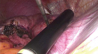
Fig. 6.2
Laparoscopic RFA
The surgical RFA approach is generally applied to patients with multiple large tumors which range from 3 to 5 cm or which are close to a large vessel. Surgical RFA allows for temporary occlusion of hepatic artery and portal vein, the Pringle’s maneuver, which practically stops all blood inflow to the liver. As the cooling effect from the blood inflow is minimized, large tumors are more likely to be completely ablated. It also facilitates bile duct cooling by using an endoscopic nasobiliary drainage (ENBD) tube during RFA for HCC close to major bile ducts. If multiple RFA ablations with overlapping ablation zones are needed for large tumors, surgical RFA is more suitable. If the patient also presents with a separate tumor that cannot undergo RFA but is suitable to be resected, it can be surgically resected during the same operation. Moreover, surgical RFA enables identification and control of bleeding after treatment of superficial tumors and avoidance of seeding along the needle track.
The disadvantages of open RFA are similar to those of other open surgeries, as it is more expensive, requires general anesthesia and longer hospital stay, and is associated with more pain. In addition, as percutaneous RFA technique has improved much in the last decade, open RFA now contributes only to a small proportion of RFA procedures in most centers. As a consequence, there are very limited data on open RFA. One non-randomized study showed percutaneous RFA to have a significantly shorter hospital stay (4.1 vs. 7.6 days) and a lower morbidity rate (2.3 % vs. 8.8 %) than open RFA [6].
Laparoscopic RFA is currently more commonly adopted than open RFA. Laparoscopic RFA combines many of the benefits of both the percutaneous and open approaches. However, a history of previous abdominal surgery with significant intraabdominal adhesions may preclude the use of the laparoscopic approach. Laparoscopic RFA is a safe treatment for liver tumors in deep locations, as well as superficial nodules adjacent to the diaphragm and organs, or for multiple lesions. The complication and mortality rates have been reported to range from 3.2 % to 27 % and from 0 % to 1.9 %, respectively [7–11]. Three non-randomized comparative studies consistently showed that laparoscopic RFA had significantly lower intraoperative blood loss, shorter operative time, and shorter postoperative hospital stay, when compared with open RFA [12–14]. In addition, laparoscopic intraoperative ultrasound during laparoscopic RFA allows a much more accurate staging than preoperative imagings. Laparoscopic intraoperative ultrasound has been reported with great accuracy during the procedure, permitting to detect 13.3–46.1 % new HCC nodules missed at preoperative imagings [15–18]. The laparoscopic approach also has the advantages of the open approach, such as applying the Pringle’s Maneuver, or to carry out concomitant resectional procedures. However, it is technically more challenging. For those tumors which are close to the dome of the liver or when the right posterior sector is shrunken in cirrhotic liver, the degree of freedom for introduction of the RFA electrodes is less than with open RFA. In expert centers, these tumors in such difficult locations can still be treated by laparoscopic RFA. In the series of caudate lobe HCC treated by laparoscopic RFA reported by the Chinese PLA General Hospital in Beijing, 27 patients underwent laparoscopic caudate lobe RFA for solitary small HCC with a mean tumor size of 2.8 cm [19]. The 1-, 3-, and 5-year overall survival rates were 96.3, 74.1, and 62.9 %, respectively. The 1-, 3-, and 5-year disease-free survival rates after RFA were 92.6, 44.4 and 33.3 %, respectively. The most common postoperative complication in that series was pleural effusion (25.9 %), followed by transient hemoglobinuria (7.4 %). For oncological outcomes, most reported series were on patients with unresectable small HCC. After a mean follow-up of 14.3–36.9 months, 2.9–69.5 % showed local recurrences; 4.8–22.3 %, remote recurrences; and <2 %, both local and remote recurrences [20–24]. Based on the limited evidence, there were no significant differences in the overall survival, recurrence-free survival, and local recurrence rates between the laparoscopic RFA group and the open RFA group. There have been more recent studies comparing liver resection with laparoscopic RFA for small HCC. Four non-randomized studies showed longer duration of hospital stay and higher rates of postoperative complications in the liver resection group [25–28]. However, the oncological outcomes were conflicting. Karabulut et al. showed no significant difference in disease-free survival between the RFA group (median tumor size, 3.1 cm) and the resection group (median tumor size, 5.3 cm), but the 5-year actual survival was significantly higher (40 % vs. 21 %) in the resection group [25]. Santambrogio et al. showed similar 5-year actuarial survival rates (54 % vs. 41 %) after resection (mean tumor size, 2.91 cm) and after RFA (mean tumor size, 2.66 cm) for solitary HCC with Child-Pugh class A liver cirrhosis, despite a marked increase in HCC recurrence rates after RFA (6 % vs. 24 %) [26]. Lai et al. showed significantly lower 5-year disease-free survival (40 % vs. 60 %) in the RFA group (mean tumor size, 1.8 cm) than in the resection group (mean tumor size, 2.9 cm) but similar 5-year overall survivals in the two groups (84 % vs. 71 %) [27]. Tohmeno et al. showed no significant differences in 5-year overall survival (35 % vs. 47 %) and 5-year disease-free survival (28 % vs. 34 %) between the RFA group (mean tumor size, 2.36 cm) and the resection group (mean tumor size, 3.07 cm) [28].
In general, surgical RFA should be considered if any of the following conditions are present: (a) significant coagulopathy, (b) large tumors (but <5 cm) or multiple lesions requiring repeated punctures, (c) superficial lesions adjacent to visceral structures, (d) lesions with a very difficult or impossible percutaneous approach, (e) short interval of recurrence of HCC following percutaneous RFA. Currently, there is no evidence which comes from randomized trial comparing percutaneous RFA and surgical RFA. All the evidence were based on cohort studies and non-randomized retrospective studies.
6.4 Conclusion
Careful patient selection and use of the best approach (percutaneous, laparoscopy, or laparotomy) help to minimize complications after RFA. The choice of the different approaches of RFA should be tailored to the individual patients according to tumor volume and location.
References
1.
2.
Kondo Y, Yoshida H, Shiina S, Tateishi R, Teratani T, Omata M. Artificial ascites technique for percutaneous radiofrequency ablation of liver cancer adjacent to the gastrointestinal tract. Br J Surg. 2006;93(10):1277–82.CrossRefPubMed
Stay updated, free articles. Join our Telegram channel

Full access? Get Clinical Tree


