| SKULL BASE REGION | Cerebellopontine angle/internal auditory canal |
| HISTOPATHOLOGY | Schwannoma |
| PRIOR SURGICAL RESECTION | Yes |
| PERTINENT LABORATORY FINDINGS | Pretreatment audiogram: Class B hearing with word recognition scores at 65% and pure tone average of 40 dB in the left ear; normal right hearing |
Case description
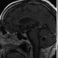
The patient was a 33-year-old woman who noted difficulty with balance and progressive left-sided facial numbness and hearing loss over several months. She underwent a brain magnetic resonance imaging (MRI) with gadolinium contrast, revealing an exceptionally large enhancing tumor in the cerebellopontine angle (CPA) ( Figure 8.39.1 ). There was associated mass effect on the pons and cerebellum with parenchymal distortion but without edema or hydrocephalus. An audiogram showed mild-to-moderate sensorineural hearing loss, with word recognition at 65% ( Figure 8.39.2 ). Neurological examination revealed mild numbness in the left V2 distribution and slight imbalance with tandem walking. She had no history of vertigo or facial weakness.


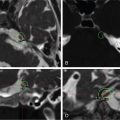
Due to the size and symptomatic nature of the lesion, as well as her young age, she underwent a left retrosigmoid craniotomy for aggressive, but subtotal, resection to preserve the facial nerve. Hearing could not be preserved. The tumor was confirmed to be a vestibular schwannoma (VS). Postoperative facial nerve function was House-Brackman (HB) grade II but returned to normal by the 3-month follow-up. She experienced altered taste involving the left tongue, as well as left nasal and ocular dryness, signifying nervus intermedius dysfunction, all of which improved at 3 months. Postoperative MRI revealed a 6-mm plaque of tumor along the facial nerve ( Figure 8.39.3 ). This residual tumor subsequently demonstrated growth at 3.5 years ( Figure 8.39.4 ), necessitating treatment with Gamma Knife radiosurgery (GKRS) ( Figure 8.39.5 ).
| Radiosurgery Machine | Gamma Knife – Perfexion |
| Radiosurgery Dose (Gy) |
|
| Number of Fractions | 1 |
Related posts:
 Esthesioneuroblastoma – delayed postoperative radiosurgery for recurrence at long-term
 Null cell – delayed postoperative radiosurgery for growing perioptic residual
 Suprasellar non-small cell lung carcinoma metastasis – upfront radiosurgery
 Chondrosarcoma – definitive radiosurgery after subtotal resections
 Trigeminal neuralgia due to microvascular conflict – upfront radiosurgery
 Capillary hemangioma – postoperative radiosurgery for residual tumor
Stay updated, free articles. Join our Telegram channel

Full access? Get Clinical Tree


