Fig. 34.1
Intraoperative photograph of colon resection for C. difficile associated disease
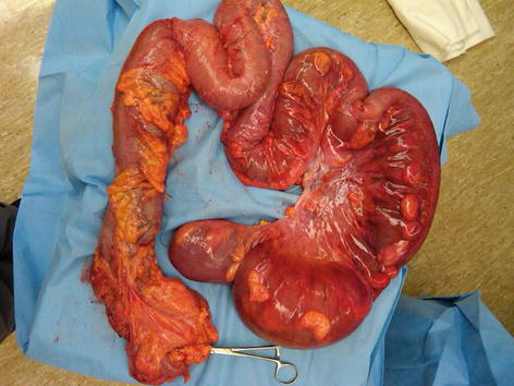
Fig. 34.2
Resected specimen of total abdominal colectomy for C. difficile induced toxic megacolon
Imaging Techniques
Abdominal imaging modalities especially plain radiographs and computed tomography (CT) are widely used to establish the cause and monitor treatment response in these patients. Plain radiograph can be quickly performed at bedside and is usually the initial modality to be used.
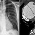
Abdominal X-Ray
Upright abdominal radiograph can detect intra-abdominal free air as little as 1–2 mm3. It is also valuable to assess the intestinal gas pattern and distinguish generalized postsurgical adynamic ileus from mechanical obstruction, although both the sensitivity and specificity are limited in many cases due to portable technique and difficulty of maintaining upright position by many ICU patients. Plain radiography can also detect large amount of ascites, pneumatosis and severe bowel wall thickening, although the sensitivity and specificity are both far inferior to CT scan. Serial radiographs are valuable to monitor the progression/resolution.
Related posts:


Stay updated, free articles. Join our Telegram channel

Full access? Get Clinical Tree


