Lemierre’s Syndrome
Shaun R. Rybak
CLINICAL HISTORY
27-year-old male with a 1-week history of sore throat, fever, and neck pain.
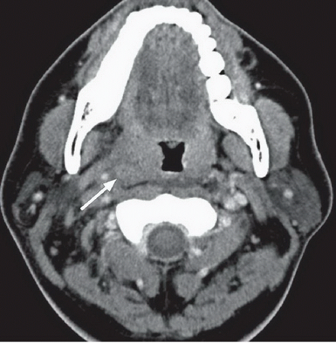 FIGURE 29A |
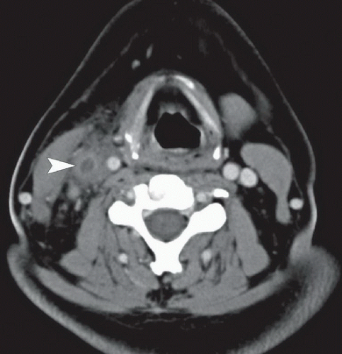 FIGURE 29B |
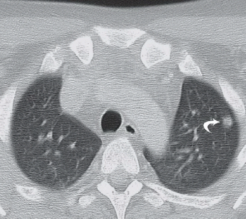 FIGURE 29C |
FINDINGS
Figure 29A: Contrast enhanced neck CT demonstrating Asymmetric enlargement of the right palatine tonsil (arrow) with adjacent parapharyngeal and carotid space fat stranding, and a small retropharyngeal effusion. Figure 29B: An oval, rim-enhancing, centrally hypodense lesion (arrowhead) with surrounding fat stranding is noted lateral to and displacing the right internal carotid artery. Note the absence of a normal contrast-filled internal jugular vein on this side. Figure 29C: There is a left upper lobe peripheral nodule with hazy margins (curved arrow). Although not shown, there were several additional similar-appearing mostly subpleural lung nodules present bilaterally.
Related posts:


Stay updated, free articles. Join our Telegram channel

Full access? Get Clinical Tree


