57 Left lobe of liver, lateral segment, subsegments II and III

58 Left lobe of liver, ligamentum teres, boundary between lateral and medial segments
The liver is divided into a left and a right lobe on anatomical criteria. The left lobe corresponds to the lateral segment; the right lobe consists of the medial, anterior, and posterior segments.
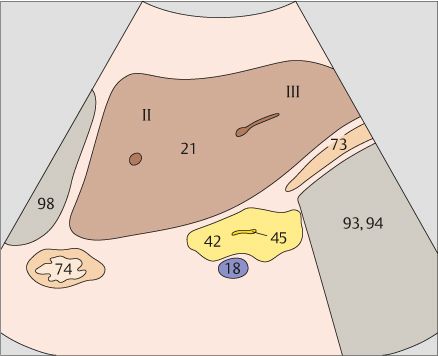
On functional criteria, the lateral and medial segments belong to the left lobe of the liver while the anterior and posterior segments belong to the right lobe.
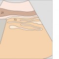
59 Left lobe of liver, ligamentum teres, boundary between lateral and medial segments
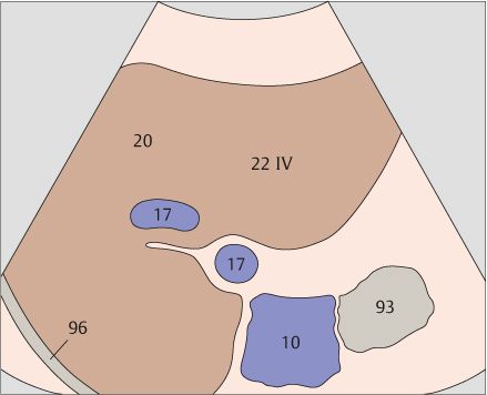
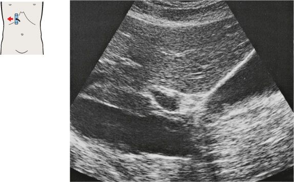
60 Left hepatic vein, ligamentum teres, boundary between lateral and medial segments, caudate lobe
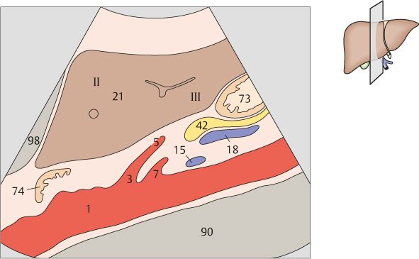
The lateral segment is composed of subsegment II cranially and subsegment III caudally.
The boundary between the lateral and medial segments, i.e., between the anatomical left and right lobes of the liver, is the left hepatic vein.
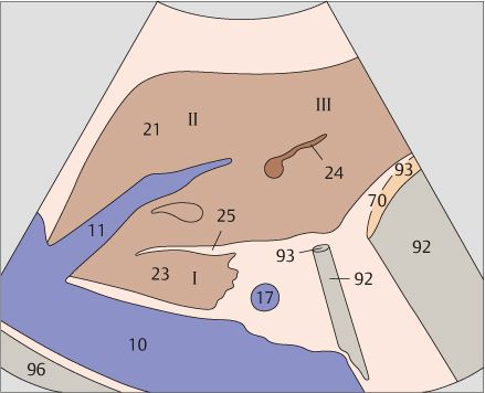
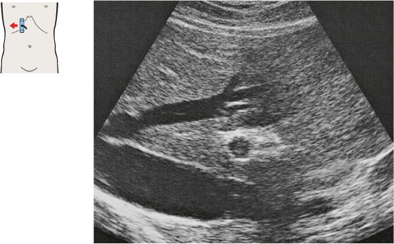
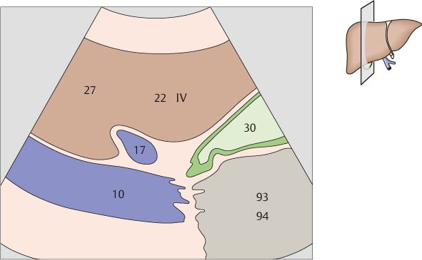
61 Left hepatic vein, ligamentum teres, boundary between lateral and medial segments, caudate lobe
62 Left hepatic vein, ligamentum teres, boundary between lateral and medial segments, caudate lobe
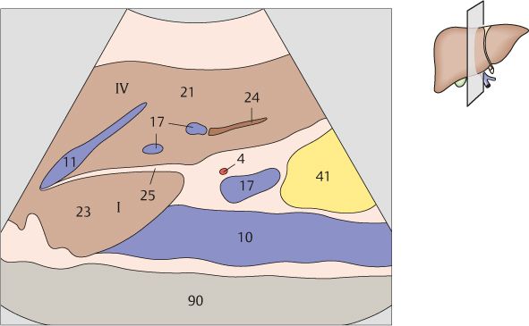
The caudate lobe corresponds to subsegment I of the medial segment and is located lateral and anterior to the vena cava. Most of the medial segment consists of subsegment IV.
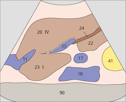
The boundary between the lateral and medial segments, i.e., between the anatomical left and right lobes of the liver, is the ligamentum teres.
63 Left hepatic vein, ligamentum teres, boundary between lateral and medial segments, caudate lobe
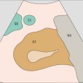
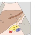
64 Medial segment, subsegment IV, quadrate lobe

Ligamentum teres (the obliterated umbilical vein) extends from the left portal vein branch to the anterior inferior border of the liver.
The caudal part of the medial segment, the quadrate lobe, is situated between ligamentum teres and the gallbladder. The quadrate lobe is part of subsegment IV.
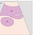
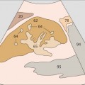
65 Gallbladder, portal vein, vena cava, boundary between medial and anterior segments
66 Middle hepatic vein, boundary between medial and anterior segments
The plane of the gallbladder and vena cava forms the boundary plane between the medial and anterior segments of the liver.
The middle hepatic vein marks the boundary between the medial and anterior segments in the cranial part of the liver.
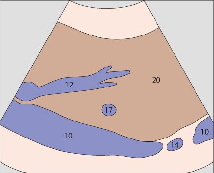
67 Anterior segment, subsegments VIII and V
68 Right hepatic vein, boundary between anterior and posterior segments
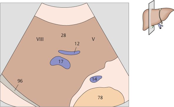
The anterior segment consists of subsegment VIII cranially and subsegment V caudally.
The right hepatic vein and the division of the right portal vein branch mark the boundary plane between the anterior and posterior segments.
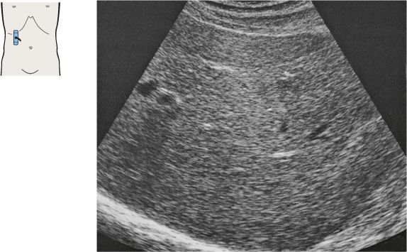
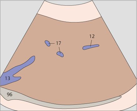
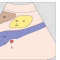
69 Posterior segment, subsegments VII and VI
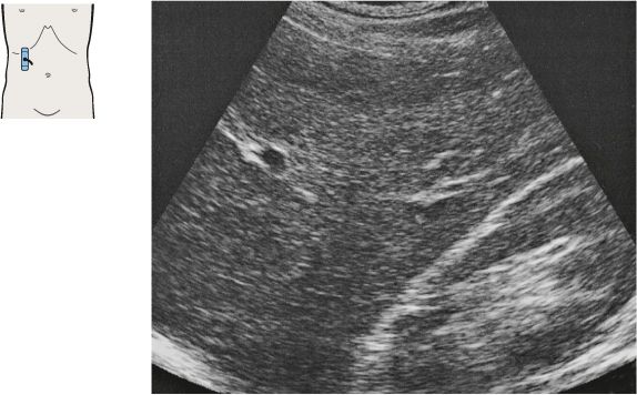
70 Posterior segment, lateral portions of liver, kidney
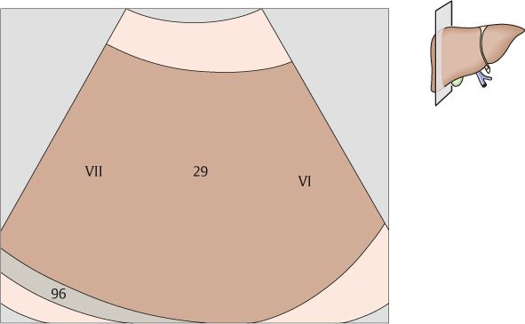
The posterior segment consists of subsegment VII cranially and subsegment VI caudally.
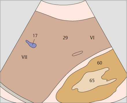
The right lobe of the liver is highly variable in its caudal extent.
Stay updated, free articles. Join our Telegram channel

Full access? Get Clinical Tree


