Liver Laceration
Kelly L. Hastings
CLINICAL HISTORY
25-year-old restrained driver in a motor vehicle collision.
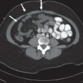
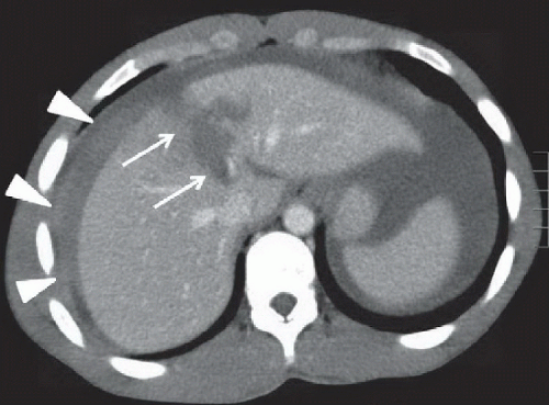 FIGURE 47A |
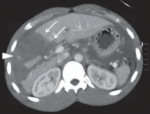 FIGURE 47B |
FINDINGS
Figure 47A: Axial contrast-enhanced CT of the liver at the level of the hepatic veins. Multiple linear and branching low-attenuation lacerations involving hepatic segments VIII and IVa extend to the vascular pedicle of the porta hepatis (arrows). Associated hemoperitoneum in the perihepatic region identified (arrowheads). Figure 47B: Contrast-enhanced CT at the level of the portal veins. Extension of the laceration into a large region of low attenuation involving most of hepatic segments V and VI (arrows). Amorphous high-density material within the large laceration/hematoma (arrowhead), representing active contrast extravasation..
DIFFERENTIAL DIAGNOSIS
Hepatic laceration, fatty infiltration, cholangiocarcinoma.
DIAGNOSIS
Related posts:


Stay updated, free articles. Join our Telegram channel

Full access? Get Clinical Tree


