Introduction
Guidelines for the use of elastography in diffuse liver disease have been released by societies or federations of societies. The first set of guidelines was released by European Federation of Societies for Ultrasound in Medicine and Biology (EFSUMB) in 2013. In 2015 the World Federation for Ultrasound in Medicine and Biology (WFUMB) and the Society of Radiologists in Ultrasound (SRU) produced guidelines on this topic, and the European Association for the Study of the Liver (EASL), together with the Asociación Latinoamericana para el Estudio del Hígado (ALEH), released clinical practice guidelines for the use of noninvasive tests in the evaluation of liver disease. The advances in the technologies and the rapidly increasing evidence in the past few years have prompted all these societies/federations of societies to update their guidelines: EFSUMB did so in 2017, WFUMB in 2018, SRU in 2020, and EASL in 2021.
All guidelines have highlighted that a correct acquisition’s protocol is necessary to obtain a liver stiffness measurement (LSM) of good quality and that the literature shows that the acoustic radiation force impulse (ARFI) techniques have similar or better accuracy than vibration controlled transient elastography (VCTE) in the assessment of liver fibrosis even though the evidence is still low. Details about the correct acquisition’s protocol are given in Chapter 4 . Guidelines have also highlighted that ultrasound (US) shear wave elastography (SWE) techniques measure liver stiffness, not fibrosis. Beside liver fibrosis, there are other factors that may lead to an increase in stiffness. These confounding factors are presented in detail in Chapter 4 .
It has been underscored that there is a large overlap of stiffness values between benign and malignant liver lesions and that there is insufficient evidence to make a recommendation on the use of SWE for differentiating and/or characterizing them. ,
For the staging of liver fibrosis in patients with chronic viral hepatitis or nonalcoholic fatty liver disease (NAFLD), the “rule of 5” has been proposed for VCTE and “rule of 4” for the ARFI techniques. , This topic is presented in detail in Chapter 7 .
European federation of societies for ultrasound in medicine and biology guidelines , ,
EFSUMB produced guidelines and recommendations on the clinical applications of US elastography in 2013; the largest section of the document was dedicated to the liver and was mainly based on the use of VCTE because the technique had been available for almost a decade at that time. In 2017 an update of the guidelines, dedicated to the clinical use of the SWE techniques for the assessment of diffuse liver disease, was released. , Based on literature data, the document evaluated the role of the SWE techniques in different etiologies of liver diseases and in several clinical scenarios. The highest level of evidence was in patients with chronic viral hepatitis, and VCTE was still the most validated technique.
The document contains several recommendations that are listed below. They were graded according to the Oxford Centre for Evidence-Based Medicine in which the level of evidence (LoE) from published studies goes from 1 (the highest level) to 5 (the lowest level). The grade of recommendation (GoR) goes from A (strongest) to D (weakest). The consensus reached through voting of the expert on each recommendation was also reported.
Recommendation 1 : The operator must acquire appropriate knowledge and training in US elastography (LoE 5, GoR C; strong consensus).
Recommendation 2 : Data acquisition should be undertaken by dedicated and specially trained personnel. For point shear wave elastography (pSWE) and two-dimensional shear wave elastography (2DSWE), experience in B-mode ultrasound is mandatory (LoE 5, GoR C; strong consensus).
Recommendation 3 : LSM by SWE should be performed through a right intercostal space in supine position, with the right arm in extension, during breath hold, avoiding deep inspiration prior to the breath hold (LoE 2b, GoR B; strong consensus).
Recommendation 4 : LSM by SWE should be performed by experienced operators (LoE 2b, GoR B; strong consensus).
Recommendation 5 : LSM by pSWE and 2D-SWE should be performed at least 10 mm below the liver capsule (LoE 1b, GoR A; strong consensus).
Recommendation 6 : Patients should fast for a minimum of 2 hours and rest for a minimum of 10 minutes before undergoing LSM with SWE (LoE 2b, GoR B; majority consensus).
Recommendation 7 : The major potential confounding factors (liver inflammation indicated by aspartate aminotransferase (AST) and/or alanine aminotransferase (ALT) elevation more than 5 times the normal limits, obstructive cholestasis, liver congestion, acute hepatitis and infiltrative liver diseases) should be excluded before performing LSM with SWE to avoid overestimation of liver fibrosis (LoE 1b, GoR B; broad consensus).
Recommendation 8 : SWE within the normal range can rule out significant liver fibrosis when in agreement with the clinical and laboratory background (LoE 2a, GoR B; broad consensus).
Recommendation 9 : For VCTE, 10 measurements should be obtained. An interquartile range/median ≤30% of the 10 measurements is the most important reliability criterion (LoE 1b, GoR A; strong consensus).
Recommendation 10 : For VCTE, values obtained with XL probe are usually lower than with the M probe, therefore no recommendation on the cutoffs to be used can be given (LoE 2b, GoR B; broad consensus).
Recommendation 11 : Adequate B-mode liver image is a prerequisite for pSWE and 2D-SWE measurements (LoE 5, GoR D; strong consensus).
Recommendation 12 : The median value of at least 10 measurements should be used for liver elastography by pSWE (LoE 2b, GoR B; strong consensus).
Recommendation 13 : For 2D-SWE, a minimum of three measurements should be obtained; the final result should be expressed as median together with interquartile range (LoE 2b, GoR B; strong consensus).
Recommendation 14 : Methods to objectively assess strain are being developed but currently cannot be recommended in clinical practice (LoE 5, GoR D; strong consensus).
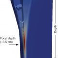
Recommendation 15 : The results with the lowest variability in comparing different pSWE or 2D-SWE systems were obtained at a depth of 4–5 cm from the transducers (with convex transducers). Accordingly, if this location is technically suitable, it would be recommended (LoE 4, GoR C; broad consensus).
Recommendation 16 : VCTE can be used as first-line assessment for the severity of liver fibrosis in patients with chronic viral hepatitis C (HCV). It performs best to rule out cirrhosis (LoE 1b, GoR A; broad consensus).
Recommendation 17 : pSWE, as demonstrated with virtual touch quantification (VTQ), can be used as first-line assessment for the severity of liver fibrosis in patients with chronic HCV. It performs best to rule out cirrhosis (LoE 2a, GoR B; broad consensus).
Recommendation 18 : 2D-SWE, as demonstrated with the SuperSonic Imagine (SSI) modality, can be used as a first-line assessment for the severity of liver fibrosis in patients with chronic HCV. It performs best to rule out cirrhosis (LoE 1b, GoR A; broad consensus).
Recommendation 19 : SWE is not recommended to monitor fibrosis changes during anti-HCV treatment (LoE 3, GoR D; strong consensus).
Recommendation 20 : LSM changes after successful anti-HCV treatment should not affect the management strategy (e.g., surveillance for hepatocellular carcinoma [HCC] occurrence in patients at risk) (LoE 3, GoR D; broad consensus).
Recommendation 21 : VCTE is useful in patients with chronic hepatitis B virus (HBV) to identify those with cirrhosis. Concomitant assessment of transaminases is required to exclude flare (elevation >5 times upper limit of normal) (LoE 1b, GoR A; broad consensus).
Recommendation 22 : VCTE is useful in carriers of inactive HBV to rule out fibrosis (LoE 2, GoR B; strong consensus).
Recommendation 23 : pSWE, as demonstrated with VTQ, is useful in patients with chronic HBV to identify those with cirrhosis (LoE 2a, GoR B; strong consensus).
Recommendation 24 : 2D-SWE, as demonstrated with SSI, is useful in patients with chronic HBV to identify those with cirrhosis (LoE 3a, GoR C; broad consensus).
Recommendation 25 : LSM changes under HBV treatment should not affect the management strategy (e.g., surveillance for HCC occurrence in patients at risk) (LoE 2b, GoR B; strong consensus).
Recommendation 26 : VCTE can be used to exclude cirrhosis in patients with NAFLD (LoE 2a, GoR B; broad consensus).
Recommendation 27 : VCTE can be used to exclude cirrhosis in patients with alcoholic liver disease, provided that acute alcoholic hepatitis is not present (LoE 2b, GoR B; strong consensus).
Recommendation 28 : LSM with VCTE is useful to identify patients with a high likelihood of having clinically significant portal hypertension (hepatic venous pressure gradient [HVPG] ≥10 mmHg) (LoE 2b, GoR B; strong consensus).
Recommendation 29 : Liver stiffness using VCTE combined with platelet count is useful to rule out varices requiring treatment. Although preliminary results are encouraging, there is insufficient evidence to recommend pSWE and 2D-SWE in this setting (LoE 2b, GoR B; broad consensus).
World federation for ultrasound in medicine and biology guidelines ,
WFUMB released the first set of guidelines for the use of elastographic techniques in 2015. They were composed of three documents: basic principles and terminology, liver elastography, and breast elastography.
The recommendations were based on the international literature and on the expertise of the team designated by WFUMB. The recommendations were not graded because at that time it was felt that there was a great heterogeneity between studies and the available evidence was still low.
An update to the liver guidelines was released in 2018, and this time the recommendations were based on published literature, and the strength of each recommendation was judged according to the Oxford Centre for Evidence-Based Medicine. The recommendations are reported below. There was strong consensus of the expert panel for all the recommendations.
Strain elastography was included in the WFUMB guidelines because this technique was still performed in some Asian countries. However, the limitations of the technique and the fact that it has an accuracy that is not good enough to diagnose any stage of liver fibrosis were also highlighted, and no specific recommendation on its use was given.
Recommendation 1 : Cutoffs for staging liver fibrosis are system specific (LoE 1b, GoR A).
Recommendation 2 : The impact of hepatic steatosis on liver stiffness is uncertain. Clinicians should exercise caution when interpreting liver stiffness results in patients with severe steatosis and obesity (LoE5, GoR C).
Recommendation 3 : SWE is useful to exclude significant fibrosis and diagnose cirrhosis in patients with untreated chronic hepatitis B (LoE1a, GoR A).
Recommendation 4 : Liver stiffness usually decreases during antiviral treatment with analogues. Screening for HCC and portal hypertension should continue despite decreased liver stiffness in patients with advanced disease (LoE1b, GoR A).
Recommendation 5 : SWE is the preferred method as the first-line assessment for the severity of liver fibrosis in untreated patients with chronic viral hepatitis C. It is useful to rule out advanced disease (LoE1a, GoR A).
Recommendation 6 : Liver stiffness decreases significantly after SVR to treatment with interferon-based therapies or direct-acting antiviral agents. However, liver stiffness cannot be used to stage liver fibrosis or rule out cirrhosis, given the loss of accuracy of cutoffs defined in viremic patients. Screening for HCC and portal hypertension should continue despite decrease of liver stiffness in patients with advanced disease (LoE1b, GoR A).
Recommendation 7 : SWE can be used for liver stiffness assessment in patients with NAFLD to rule out advanced fibrosis and select patients for further assessment (LoE1a, GoR A).
Recommendation 8 : SWE can be used for liver stiffness assessment in patients with ALD to rule out advanced disease. Caution is needed in patients with ongoing alcohol abuse or with acute alcoholic hepatitis (LoE 2a, GoR B).
Recommendation 9 : SWE has high diagnostic accuracy for detecting cirrhosis; better at ruling out (high NPV above 90%) than ruling in (LoE 1a, GoR A).
Recommendation 10 : LSM of VCTE >20 kPa can be used to identify patients likely bearing clinically significant portal hypertension (HVPG ≥10 mmHg) (LoE 2b, GoR B).
Recommendation 11 : LSM using VCTE <20–25 kPa combined with platelet count >110,000–150,000/μL is useful to rule out varices needing treatment (LoE 2b, GoR B).
Recommendation 12 : LSM holds prognostic value in compensated cirrhosis and the higher the value, the higher the risk of clinical complications (LoE 2b, GoR B).
Recommendation 13 : There is insufficient evidence to make a recommendation on the use of SWE for liver stiffness assessment in pediatric patients (LoE 5, GoR D).
Recommendation 14 : There is insufficient evidence to make a recommendation on the use of SWE for differentiation between benign and malignant lesions and characterization of focal liver lesions (LoE 5, GoR D).
Recommendation 15 : Controlled attenuation parameter is a point of care, standardized and reproducible technique, promising for the detection of liver steatosis. However, for quantifying steatosis, there is a large overlap between adjacent grades, there are no consensual cutoffs, and quality criteria are not well defined (LoE 3, GoR C).
Recommendation 16 : Interpretation of liver stiffness measurements needs to be taken in context with the other clinical and laboratory data (LoE 1b, GoR A).
Society of radiologists in ultrasound consensus , ,
In 2014 the SRU convened a panel of specialists from radiology, hepatology, pathology, and basic science and physics to arrive at a consensus regarding the use of elastography in the assessment of liver fibrosis in chronic liver disease. The results of the panel were published in 2015. ,
The conference statement discussed the burden of chronic liver disease and cirrhosis, predisposing conditions, and the staging of fibrosis. The panel concluded that the histologic reference standard was imperfect with high variability between pathologist. It listed the sources of variability in elastography: (1) origin of underlying disease, (2) patient comorbidities, (3) modality being used, (4) system-specific factors, (5) machine variability, (6) patient physical factors, (7) indication for study, (8) disease prevalence, (9) patient sex, (10) postprandial state, and (11) breath-hold technique. The panel provided a comparison of the elastographic modalities including advantages, expenses, frequency of shear wave generation, limitations, measurement location, region of interest size, value reported, and defining a good measurement.
The panel provided evidence-based best practice for performance of US-based elastography. These included fasting for 4–6 hours, specific positioning of patient, right arm elevated above the head, shallow breath hold, placement of the region of interest, and the acquisition number of measurements.
A review of the pSWE, 2D-SWE, VCTE, and magnetic resonance elastography (MRE) techniques were provided, highlighting the variability of the measurements in noninvasive assessment of liver fibrosis. Due to the large overlap of stiffness values in intermediate levels of fibrosis regardless of which technique was used, a low cutoff value for various systems was provided, below which there was no clinically significant fibrosis and a high cutoff value, above which there was a high probability of compensated advanced chronic liver disease (cACLD). It was recommended that using likelihood ratios was more appropriate than converting the stiffness values in METAVIR scores. A recommendation of what should be included in the report was provided.
In 2020 the SRU published an update to the SRU liver elastography consensus statement. The update again emphasized following the strict protocol recommended in the 2015 document. The experts panel underscored that most patients with HCV are being treated regardless of the degree of fibrosis. Because the overlap of liver stiffness values between METAVIR scores is as large if not larger than the difference between vendors, separate cutoff values for each vendor were not required. It was pointed out that what is most clinically important is diagnosing cACLD. Given the large overlap of stiffness values for mild to moderate fibrosis, the SRU continued to recommend a low cutoff value, below which there is a high probability of no or mild fibrosis and recommended a high cutoff value, above which there is a high probability of cACLD. The recommendations made by the panel are discussed in detail in Chapter 7 . Briefly, a vendor neutral “rule of 4” for interpretation of liver stiffness values obtained using ARFI techniques in patients with viral hepatitis and NAFLD was proposed. In patients with chronic HBV or HCV hepatitis successfully treated with antiviral drugs, the cutoffs obtained in treatment-naive patients should not be used because a rapid decline of stiffness values is observed in treated subjects, likely due to the decrease of liver inflammation. Therefore, when liver cirrhosis is evident by B-mode findings, elastography should not be used to rule out the disease because a value in the low range of liver stiffness may only indicate a successful response to antiviral treatment. The panel suggested that the delta change of liver stiffness values over time should be used instead of the absolute values to follow-up patients. Thus every patient becomes their own control. In patients with chronic viral hepatitis who are successfully treated, the baseline liver stiffness value to be used for the follow up should be that obtained after viral eradication or suppression.
For pediatric patients with liver disease or congenital heart disease with Fontan surgery, it was recommended that each subject becomes their own control, using the stiffness delta changes over time to evaluate the efficacy of the treatment or the progression of disease.
The consensus points out that by applying this rule, liver stiffness assessment can be suitable for evaluating all clinical conditions that increase liver stiffness, independent of the disease etiology, including nonfibrotic causes of liver stiffness increase, such as congestive heart failure.
Because there is an approximately 10% variability of the measurements within a vendor and between vendors, the recommendation is that a clinically significant change should be considered when the delta is greater than 10%. The panel recommended using the same equipment for follow-up studies.
Asian-pacific association for the study of the liver guidelines
In 2016 the Asian-Pacific Association for the Study of the Liver (APASL) guidelines developed consensus guidelines on the assessment of liver fibrosis that included invasive methods, such as liver biopsy and hepatic venous pressure gradient measurements, and noninvasive methods, such as biomarkers, conventional radiological methods, and elastography techniques. In these guidelines, liver biopsy was considered the gold standard; however, all the limitations of the procedure were highlighted. In the short paragraph dedicated to elastography techniques, it was acknowledged that ARFI-based methods are as accurate as VCTE in staging liver fibrosis.
American gastroenterological association guidelines
The American Gastroenterological Association (AGA) guidelines, published in 2017, were focused on the role of VCTE for the evaluation of liver fibrosis. At that time there was already an abundant literature on this topic, including several metaanalyses; however, all the recommendations were graded as conditional and with low-quality evidence. In the AGA guidelines it is claimed that VCTE is the most commonly used imaging-based technique for fibrosis assessment method in the United States; however, no evidence is provided, thus this statement could be misleading. Moreover, the term elastography is identified with VCTE, and there is no mention of the ARFI techniques or to the literature data showing that these techniques have an accuracy that is at least similar to that reported for VCTE.
European association for the study of the liver guidelines ,
The first set of EASL guidelines was produced together with the ALEH in 2015. It indicates that, for fibrosis staging, VCTE should be considered the reference standard because it is the most validated. Nonetheless, the EASL–ALEH guidelines acknowledged that the pSWE and 2D-SWE techniques showed the same accuracy of VCTE for the staging of liver fibrosis in published studies, even though the level of available evidence was lower than that of VCTE. This trend has been maintained in the recently released update.
In the update, the noninvasive tests that were analyzed for the evaluation of liver disease severity and prognosis were blood markers, methods assessing physical properties of the liver tissue (e.g., liver stiffness, attenuation, viscosity), and imaging methods assessing the anatomy of the liver and other abdominal organs (e.g., US, MR, and CT).
The panel of experts developed the clinical practice guidelines (CPGs) update according to a format based on P: Patient, Population, or Problem; I: Intervention, Prognostic Factor, or Exposure; C: Comparison or Intervention (if appropriate); and O: Outcome questions. The CPGs were sent to a Delphi Panel for reviewing and voting. The suggested changes were taken into consideration in a revised version, which was then submitted to the EASL Governing Board. The following scenarios were analyzed: general population; alcohol-related liver disease; HCV postsustained virological response (SVR)/postantiviral therapy; NAFLD/nonalcoholic steatohepatitis (NASH); cholestatic and autoimmune liver disease; cACLD, and portal hypertension. The LoE based on the Oxford Centre for Evidence-Based Medicine and the QUADAS-2 tool for accuracy of diagnostic studies were used to judge the quality of the evidence.
General population
It is underscored that, because of the spectrum effect, noninvasive fibrosis tests will likely have lower sensitivity and higher specificity in populations with lower prevalence of the disease.
Noninvasive tests should be preferentially used in patients at risk of advanced fibrosis, such as patients with metabolic risk factors and/or harmful use of alcohol, and not in unselected populations (LoE 2). They should be used to rule out rather than rule in advanced fibrosis (LoE 1). ALT, AST, and platelet count should be part of the routine investigations in primary care in patients with suspected liver disease so that simple non invasive scores can be readily calculated (LoE 2). The automatic calculation and systematic reporting of simple non invasive fibrosis tests such as the fibrosis-4 index for liver fibrosis (FIB-4) in populations at risk of liver fibrosis (individuals with metabolic risk factors and/or harmful use of alcohol) in primary care is recommended to improve risk stratification and linkage to care (LoE 2). In low-prevalence populations, all noninvasive tests can identify advanced fibrosis in patients at risk significantly better than clinical acumen alone (LoE 1).
Individuals at risk for advanced fibrosis should be entered in appropriate risk stratification pathways using noninvasive fibrosis tests (LoE 1). In low-prevalence populations the choice of noninvasive tests and the design of diagnostic pathways should be performed in consultation with a liver specialist (LoE 3).
All these recommendations were graded as strong.
Alcohol-related liver disease
Accuracy of noninvasive methods for fibrosis staging compared to liver biopsy
The followings are strong recommendations:
- ■
Liver stiffness values by VCTE <8 kPa can rule out alcohol-related advanced chronic liver disease (ACLD) (LoE 3); values ≥12–15 kPa can diagnose ACLD in high-prevalence populations only after excluding false positive cases (LoE 2).
- ■
In patients with elevated liver stiffness and evidence of hepatic inflammation by blood tests (AST or gamma-glutamyl transferase higher than two times the upper limit of normal), liver stiffness by VCTE should be repeated after at least 1 week of alcohol restraint (LoE 3).
Accuracy of noninvasive tests for predicting liver-related outcomes compared to liver biopsy, HVPG, child-pugh score, or model for end-stage liver disease
Because of lack of evidence, the panel felt that was not possible to make any recommendations regarding prognostic markers in alcohol-related compensated liver disease.
HCV post-SVR/postantiviral therapy
Accuracy of noninvasive tests compared to liver biopsy to stage liver fibrosis in patients with cACLD who achieved SVR after antiviral therapy
- ■
The routine use of noninvasive scores and liver stiffness is not recommended because they lack accuracy in detecting fibrosis regression (LoE 3).
- ■
Liver stiffness cutoffs that were obtained in untreated patients with HCV should not be used to stage liver fibrosis after SVR (LoE 4).
These were strong recommendations.
Accuracy of noninvasive tests to predict clinical outcomes (decompensation, HCC) compared to liver biopsy, HVPG, child-pugh score or MELD in patients with HCV and patients with cACLD who achieved SVR
In patients with cACLD previous to antiviral therapy for HCV, LSM post-SVR could be helpful to refine the stratification of residual risk of liver-related complications and liver stiffness can be repeated yearly (LoE 3). Patients with cACLD prior to treatment should continue to be monitored for HCC and portal hypertension regardless of the results of noninvasive tests post-SVR (LoE 3; strong recommendation).
NAFLD/NASH
Accuracy of noninvasive tests for the diagnosis of steatosis compared to liver biopsy
The studies performed using serum scores or controlled attenuation parameter were evaluated. The conclusion was that noninvasive scores are not recommended for the diagnosis of steatosis in the clinical practice (LoE 2; strong recommendation), whereas conventional US is recommended as a first-line diagnostic tool despite its well-known limitations (LoE 1; strong recommendation).
Magnetic resonance imaging-estimated proton density fat fraction (MRI-PDFF) is the most accurate noninvasive method for detecting and quantifying steatosis; however, it is not recommended as a first-line tool due to its cost and limited availability (LoE 2; strong recommendation).
Accuracy of noninvasive tests to evaluate NAFLD severity (presence of NASH and staging of liver fibrosis) compared to liver biopsy
It is highlighted that liver biopsy is still the reference standard for the diagnosis of NASH, because none of the available noninvasive tests has acceptable accuracy (LoE 2).
In patients with NAFLD, the following tests are recommended to rule out advanced fibrosis in clinical practice (LoE 1, strong recommendation): liver stiffness by VCTE <8 kPa; patented tests: ELF <9.8 or FibroMeter <0.45 or FibroTest < 0.48; nonpatented tests: FIB-4 <1.3 or NAFLD fibrosis score < −1.455. In patients with FIB-4 >1.3, VCTE and/or patented serum tests should be used to rule out or rule in advanced fibrosis (LoE 2; strong recommendation). Even though MRE is the most accurate noninvasive method for staging liver fibrosis, it is only marginally better than other noninvasive tests for the diagnosis of ACLD; therefore it is not recommended as first-line diagnostic test also due to its cost and limited availability (LoE 2; strong recommendation). It can play a role in clinical trials.
Accuracy of noninvasive tests to predict liver-related outcomes in patients with NAFLD compared to liver biopsy, HVPG, child-pugh score, or MELD
Serum scores and liver stiffness by VCTE should be used to stratify the risk (LoE 3; strong recommendation). Repeated measurements of noninvasive tests can be used to refine stratification of risk of liver-related events in patients with NAFLD/NASH. Even though there is lack of evidence for choosing the optimal interval for the follow-up, the panel deemed it reasonable to repeat the tests every 3 years in patients in early stage of liver fibrosis and every year in patients with ACLD (LoE 3; weak recommendation).
Accuracy of noninvasive tests for patients’ selection and evaluation of treatment response in NAFLD therapeutic trials compared to liver biopsy
It was strongly recommended to use liver biopsy for the selection of patients in therapeutic trials and to evaluate NASH resolution and liver fibrosis improvement in this setting. A weak recommendation was given for the use of MRI-PDFF for assessing steatosis changes under treatment because it was felt that the relevant minimal decrease in MRI-PDFF needed to be better defined.
Cholestatic and autoimmune liver disease
Accuracy of noninvasive tests to assess disease severity in comparison to liver biopsy in patients with primary biliary cholangitis and primary sclerosing cholangitis
Noninvasive scores and serum markers are not recommended for fibrosis staging in clinical practice (LoE 3; strong recommendation).
In patients with PBC, a cutoff of 10 kPa by VCTE can rule in cACLD (LoE 3; strong recommendation).
In patients with PSC, a value of liver stiffness >9.5 kPa by VCTE can be used to support the diagnosis of advanced fibrosis in compensated patients with normal bilirubin and without high-grade stenosis (LoE 3; weak recommendation).
Accuracy of noninvasive tests to predict liver-related outcomes in patients with PBC and PSC compared to liver biopsy, HVPG, child-pugh score, or MELD
In patients with PBC, noninvasive discrimination of early and advanced stage disease based on biochemical parameters (normal vs. abnormal albumin and bilirubin) and LSM by VCTE < or >10 kPa is recommended at baseline (LoE 3; strong recommendation). During treatment, risk stratification should be based on the assessment of response to therapy by using continuous (GLOBE and UK-PBC risk scores) and/or qualitative criteria (Paris II, Toronto, Rotterdam, Barcelona, Paris I) and liver stiffness by VCTE (LoE 3; strong recommendation).
In patients with PSC, both the ELF score and liver stiffness by VCTE correlate with outcomes and they should be used for risk stratification both at baseline and during follow-up (LoE 3; strong recommendation). MRI (alone or combined with VCTE values) can be used for prognostic purposes (LoE 3; strong recommendation).
Accuracy of noninvasive test to assess liver fibrosis, and to monitor disease course as compared to liver biopsy in patients with autoimmune hepatitis
Liver stiffness by VCTE can be used in treated patients to monitor the disease course together with transaminases and immunoglobulin G and to stage liver fibrosis after at least 6 months of immunosuppressive therapy (LoE 3; weak recommendation).
Compensated advanced chronic liver disease and portal hypertension
Accuracy of noninvasive tests to diagnose cACLD as compared to liver biopsy
- ■
Patented serum tests or SWE techniques for the diagnosis of cACLD should be used in specialized settings (LoE 2; strong recommendation).
- ■
Patented serum tests (FibroTest, FibroMeter, and ELF) should be used to rule out cACLD if available (LoE 3; strong recommendation).
- ■
A cutoff <8–10 kPa by VCTE rules out cACLD, whereas a cutoff >12–15 kPa rules in the disease. Intermediate values require further testing (LoE 3; strong recommendation).
- ■
pSWE and 2D-SWE should be used to rule out and diagnose cACLD. They have shown AUROCs >0.90 in published metaanalyses (LoE 2; strong recommendation).
- ■
Intersystem variability should be taken into consideration in the interpretation of results obtained with different SWE techniques because values, ranges, and cutoffs are not comparable (LoE 3; strong recommendation).
Accuracy of noninvasive tests to diagnose clinically significant portal hypertension and to monitor portal hypertension in cACLD in comparison to HVPG measurement
- ■
A cutoff of >20–25 kPa by VCTE should be used to diagnose CSPH in patients with cACLD (LoE 1; strong recommendation).
- ■
Platelet count, spleen size, and spleen stiffness should be used as additional noninvasive tests to further improve risk stratification for CSPH (LoE 3; strong recommendation).
- ■
The presence of portosystemic collaterals on US, CT, or MR is a sign of CSPH in patients with cACLD and should be routinely reported (LoE 2; strong recommendation).
- ■
For an exact assessment of the severity of portal hypertension beyond the detection of CSPH and for assessment of the hemodynamic response to treatment, HVPG remains the only validated tool and should not be substituted by noninvasive tests (LoE 1; strong recommendation).
Accuracy of noninvasive tests to diagnose and exclude high-risk gastroesophageal varices in comparison to endoscopy
- ■
In patients with cACLD due to untreated viral hepatitis, HIV–HCV coinfection, alcohol, NAFLD, PBC, and PSC, a liver stiffness by VCTE <20 kPa and platelet count >150,000 /L (Baveno VI criteria) is a validated tool to rule out high-risk varices and avoid endoscopic screening. These criteria should be used whenever VCTE is available (LoE 1a; strong recommendation).
- ■
Spleen stiffness can be used as an additional tool to refine the risk of high-risk varices in cACLD (LoE 2; weak recommendation).
- ■
CT should not be used for primary screening for esophageal and gastric varices; however, in a routine CT, varices should be reported if present (LoE 3; strong recommendation).
Accuracy of noninvasive tests to predict clinical decompensation, hepatocellular carcinoma, and mortality in cACLD as compared to liver biopsy, HVPG, child-pugh score, or MELD
- ■
Liver stiffness at diagnosis should be used in addition to liver function tests to stratify the risk of clinical decompensation and mortality (LoE 1; strong recommendation).
- ■
Annual repeated measurements of liver stiffness can be used to refine risk stratification (LoE 5; weak recommendation).
- ■
Liver stiffness can be used in addition to clinical variables and accepted risk scores to stratify the risk of HCC in patients with HBV (LoE 3; weak recommendation).
Related posts:


Stay updated, free articles. Join our Telegram channel

Full access? Get Clinical Tree


