Fig. 1
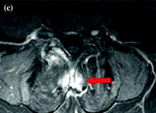
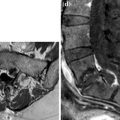
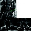
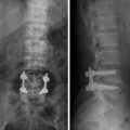
a–c. XR lateral (a) CE fat sat SE T1 sagittal (b) and axial (c). Interspinous kissing is visualized in XR (a) and MR (b). b–c MR: CE of the spinous processes and of surrounding soft tissue due to edema following reduction of amplitude of interspinous space
< div class='tao-gold-member'>
Only gold members can continue reading. Log In or Register to continue
Related posts:
Stay updated, free articles. Join our Telegram channel

Full access? Get Clinical Tree


