Lung Abscess
Sam A. Glaubiger
CLINICAL HISTORY
41-year-old female with malaise and cough.
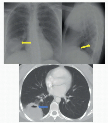 FIGURE 38A |
FINDINGS
Figure 38A: Posteroanterior (PA) plain film of the chest (left). There is a dense round opacity (yellow arrow) in the right lung base containing a small air-fluid level. Axial CT image of the chest (right). The spherical opacity is in the right lower lobe, and the air-fluid level (blue arrow) is again noted.
DIFFERENTIAL DIAGNOSIS
Lung abscess, empyema, bronchogenic carcinoma, pulmonary metastasis, diaphragmatic hernia.
DIAGNOSIS
Related posts:


Stay updated, free articles. Join our Telegram channel

Full access? Get Clinical Tree


