and Clarisse Dromain2
(1)
Department of Radiology, San Giovanni Hospital, Roma, Italy
(2)
Department of Radiology, Institut de Cancerologie Gustav Roussy, VilleJuif – Paris, France
Abstract
Malabsorption is defined as deficient absorption of any essential food materials within the small bowel. It can be primary or secondary. Celiac disease is the most common cause of primary malabsorption. Possible origin of secondary malabsorption consisted in a wide spectrum of diseases: parasites infections (Giardia, etc.), inflammatory condition (enteritis viral, bacterial, fungal, chronic inflammatory bowel disease), mechanical defects (fistulas), neurologic disorder (functional diarrhea in diabetes), endocrine disease (Zollinger–Ellison syndrome), drugs (cathartics), systemic disease (scleroderma, lupus, polyarteritis, amyloidosis), lymphoma, benign and malignant bowel tumors, pancreatic disease (pancreatitis, pancreatectomy, cystic fibrosis, pancreatic cancer), and hepatobiliary disease (biliary obstruction, chronic liver disease).
Malabsorption
Malabsorption is defined as deficient absorption of any essential food materials within the small bowel. It can be primary or secondary.
Celiac disease is the most common cause of primary malabsorption. Possible origin of secondary malabsorption consisted in a wide spectrum of diseases: parasites infections (Giardia, etc.), inflammatory condition (enteritis viral, bacterial, fungal, chronic inflammatory bowel disease), mechanical defects (fistulas), neurologic disorder (functional diarrhea in diabetes), endocrine disease (Zollinger–Ellison syndrome), drugs (cathartics), systemic disease (scleroderma, lupus, polyarteritis, amyloidosis), lymphoma, benign and malignant bowel tumors, pancreatic disease (pancreatitis, pancreatectomy, cystic fibrosis, pancreatic cancer), and hepatobiliary disease (biliary obstruction, chronic liver disease).
MALT Lymphoma
MALT lymphoma is a form of malignant B-cell non-Hodgkin’s lymphoma (MNHL) involving the mucosa-associated lymphoid tissue. It is associated with a Helicobacter pylori bacterial infection and chronic inflammation. This is a low-grade lymphoma that grows slowly and remains confined to one organ for a relatively long time.
It accounts for only about 5 % of all non-Hodgkin’s lymphomas. The most commonly affected organ is the stomach. Most gastric MALT lymphomas arise in patients in whom gastroscopy shows nonspecific features such as inflammation, superficial erosions, or ulceration. That is why the most frequent problem in the diagnosis of gastric MALT lymphoma is its differentiation from H. pylori-associated gastritis.
On CT imaging, the MALT lymphoma often appears as a thickening of the stomach wall that is focal and moderate. This finding highlighted the necessity to have a good distension with water during CT examination.
The diagnosis is made by biopsy at the time of endoscopy. Simultaneous tests for H. pylori are also done to detect the presence of this bacterium.
MALT lymphomas remain localized in the majority of cases. However, 25–35 % of MALT lymphomas, more frequently among nongastric than gastric cases, present with disseminated disease. Locoregional staging of gastric MALT lymphoma is conducted by endoscopic ultrasonography and CT. An increasing depth of invasion of lymphoma through the stomach wall, depicted using endoscopic ultrasonography, has been reported to be closely correlated with decreasing responsiveness to H. pylori eradication treatment. Similarly, the involvement of the regional lymph nodes (which occurs in approximately 15–30 % of gastric MALT lymphomas) is associated with considerably reduced response rates to either antibiotics or conventional treatment. The bone marrow is involved in this lymphoma in up to 15 % of cases, and less than 10 % of patients have distant lymph node involvement.
Treatment is tailored to organ involvement and stage at presentation. Eradication of Helicobacter pylori using a triple anti-H. pylori regimen is standard therapy for all Helicobacter pylori-positive gastric MALT lymphomas. FDG PET could be useful to follow MALT lymphoma under treatment.
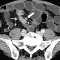
Meckel Diverticulum
A Meckel diverticulum, a true congenital diverticulum, is a small bulge in the small intestine present at birth. It is a vestigial remnant of the omphalomesenteric duct.
Meckel diverticulum is located in the distal ileum, usually within about 60–100 cm (2 ft) of the ileocecal valve. This blind segment or small pouch is about 3–6 cm long and may have a greater lumen diameter than that of the ileum. Heterotopic rests of the gastric mucosa and pancreatic tissue are seen in 60 and 6 % of cases, respectively.
A memory aid is the rule of 2s: 2 % (of the population), 2 ft (from the ileocecal valve), 2 in. (in length), 2 % are symptomatic, 2 types of common ectopic tissue (gastric and pancreatic), 2 years is the most common age at clinical presentation, and 2 times more boys are affected. The majority of people afflicted with Meckel diverticulum are asymptomatic. If symptoms do occur, they typically appear before the age of 2.
The most common presenting symptom is painless rectal bleeding such as melena-like black offensive stools, followed by intestinal obstruction, volvulus, and intussusception. Occasionally, Meckel diverticulitis may present with all the features of acute appendicitis.
A technetium-99m (99mTc) pertechnetate scan, also called Meckel scan, is the investigation of choice to diagnose Meckel diverticula. This scan detects gastric mucosa since approximately 50 % of symptomatic Meckel diverticula have ectopic gastric or pancreatic cells contained within them; this is displayed as a spot on the scan distant from the stomach itself. This scan is highly accurate and noninvasive, with 95 % specificity and 85 % sensitivity.
Computed tomography (CT scan) might be a useful tool to demonstrate a blind-ended and inflamed structure in the mid-abdominal cavity, which is not an appendix.
Megacolon
Two clinical forms of megacolon are possible: aganglionic megacolon (see Hirschsprung disease) and toxic megacolon.
Toxic megacolon is an acute transmural fulminant colitis with neurogenic loss of motor tone and rapid development of extensive colonic dilatation (>5.5 cm in transverse colon). Toxic megacolon represents commonly a complicating condition of ulcerative colitis; however, it can also be observed in patients with Crohn’s disease, salmonellosis, pseudomembranous colitis, and ischemic colitis. Clinical features of toxic megacolon are represented by systemic toxicity and profuse bloody diarrhea with colonic ileus at X-rays (marked dilatation of transverse colon with air–fluid levels) and increasing caliber of the colon on serial radiographs).
At CT toxic megacolon appears with distended colon with large amount of fluids and air, lack of colon or rectal obstruction, irregular nodular contour of thin colonic wall, and possible intramural air collection (pneumatosis).
Mesenteric Cyst
Mesenteric or omental cyst can result as a sequelae of mesenteric of omental hematoma or abscess (thick wall, internal septa) and otherwise can be the manifestation of a duplication cyst (unilocular with thin wall). In differential diagnosis pancreatic pseudocyst and cystic lymphangioma should be considered as well.
< div class='tao-gold-member'>
Only gold members can continue reading. Log In or Register to continue

Stay updated, free articles. Join our Telegram channel

Full access? Get Clinical Tree


