Magnetic resonance cholangiopancreatography (MRCP) is an elegant MR technique for noninvasively delineating the biliary system. Technologic advances in MRCP acquisition and processing and the routine availability of three-dimensional sequences have facilitated detailed assessment of biliary anatomy and pathologic or congenital processes; therefore, invasive endoscopic retrograde cholangiopancreatography is rarely needed for establishing a diagnosis. MRCP can be combined with contrast-enhanced MR imaging to enable concurrent evaluation of organs such as the liver and pancreas in addition to functional biliary imaging. This review focuses on the current use of MRCP to evaluate nonmalignant processes affecting the biliary system.
Normal anatomy and congenital variations
Knowledge of biliary anatomy is often needed to plan surgical intervention.
Bile duct anatomy and variants can be well evaluated using a standard two-dimensional (2D) magnetic resonance cholangiopancreatography (MRCP) technique. Obscuration of duct anatomy caused by overlap from adjacent fluid-filled ducts and bowel was once a limitation inherent to the standard 2D technique. However, with the routine use of three-dimensional (3D) MRCP sequences and processed maximum intensity projection (MIP) images, this problem is less often encountered.
The classically described normal anatomic configuration of the bile duct, consisting of 2 right segmental hepatic ducts (anterior and posterior) joining to form the main right hepatic duct and 2 major segmental branches of the left hepatic duct (medial and lateral) joining to form the main left hepatic duct, is present in only 50% to 60% of the population. The main right and left hepatic ducts typically fuse to form the common hepatic duct 1 cm beyond the liver margin. The common hepatic duct extends from the confluence of the right and left main ducts and becomes the common bile duct (CBD) at the point where the cystic duct inserts. The sphincter of Oddi consists of smooth muscle surrounding the common channel of the distal CBD and pancreatic duct as they insert into the duodenal papilla. The average CBD diameter is reported as 5 mm in individuals less than 50 year old; it can increase by 1 mm per decade after age 50 years. However, after cholecystectomy, the CBD is often capacious and can measure up to 13 mm, but it shows lack of intrahepatic biliary ductal dilation—a finding useful in discrimination of CBD obstruction from normal expected dilation in these patients.
Because variant biliary duct anatomy is common, it is important to identify such variants for surgical planning before major hepatectomy, liver transplantation, and laparoscopic cholecystectomy to facilitate planned liver resection and ductal anastamosis as well as to minimize inadvertent biliary injuries during surgery.
A crossover anomaly is characterized by drainage of the right posterior segmental hepatic duct into the left main hepatic duct ( Fig. 1 ). The crossover anomaly is found in 13% to 19% of the population, and is similar in prevalence to trifurcation of the biliary confluence that drains into the common hepatic duct. An accessory or aberrant right hepatic biliary duct draining into the common hepatic or cystic duct can be seen in 7.4% of the population.

The biliary ductal variants of surgical interest ( Tables 1 and 2 ) involve those that affect the outflow, length, and course of the intra- and extrahepatic biliary ducts. A crossover anomaly and trifurcation of the biliary confluence are of surgical importance when planning a left hepatectomy, because the inadvertent surgical ligation of these variant ducts leads to atrophy and cirrhosis of liver segments VI to VII or V to VIII. In addition, inadvertent resection or ligation of these anomalous ducts can lead to biliary leaks and strictures, especially in laparoscopic cholecystectomy or living donor right-lobe liver transplantation.
| Variant | Prevalence (%) |
|---|---|
| Trifurcation of the biliary duct | 19 |
| Right posterior segmental hepatic duct draining into the left hepatic duct (crossover anomaly) | 13–19 |
| Right hepatic duct emptying into the common hepatic or cystic duct | 7.4 |
| Variant | Prevalence (%) |
|---|---|
| Low medial insertion of the cystic duct into the distal CBD | 9 |
| Long parallel course of the cystic duct | 10 |
Cystic duct anomalies are correctly identified with preoperative imaging in about 18% to 23% of cases, but such identification is important to reduce risk of major injury to the biliary tree during laparoscopic cholecystectomy. For example, in patients with a low cystic duct insertion ( Fig. 2 ), the cystic duct has a long parallel course before inserting into the distal one-third of the CBD. Failure to recognize a low cystic duct insertion may lead to injury to the common duct during laparoscopic cholecystectomy. An aberrant right hepatic duct joining the common hepatic duct or cystic duct can also be inadvertently injured or ligated during laparoscopic cholecystectomy, leading to atrophy of segments VI and VII of the right hepatic lobe. Similarly, failure to recognize a proximal cystic duct insertion may lead to inadvertent ligation of the cystic duct and subsequent development of a stricture in the common hepatic duct. Long-term complications associated with low insertion of the cystic duct include postcholecystectomy syndrome, which is caused by development of calculi and inflammatory changes of a long cystic duct remnant, and instrumentation injury during endoscopic retrograde cholangiopancreatography (ERCP).

Congenital Abnormalities of the Biliary Duct
Bile duct cysts (or choledochal cysts) are rare cystic dilations of the biliary tree. There are 5 types of biliary cysts according to the Todani classification system:
- 1.
Type I choledochal cysts are the most common, comprising 80% to 90% of bile duct cysts, and are defined as fusiform dilatation of the extrahepatic CBD.
- 2.
A type II cyst is a true saccular diverticulum from the extrahepatic bile duct or an intrahepatic bile duct.
- 3.
A type III cyst, or choledochocele, represents a focal protrusion of a dilated segment of the distal CBD into the duodenum. An individual with a type III cyst may present with abdominal pain, jaundice, and vomiting, but many are incidentally detected.
4. Type IV cysts are subdivided into 2 subtypes: IVa, which shows fusiform dilation of the entire extrahepatic bile duct with extension into the intrahepatic ducts ( Fig. 3 ), and IVb, which are characterized by multiple cystic dilatations of only the extrahepatic bile duct.

Fig. 3
Type 4A choledochal cyst. A type 4A choledochal cyst ( arrow ), which represents fusiform dilation of the entire extrahepatic bile duct with extension into the intrahepatic ducts, is seen on this coronal MIP image from MRCP.
5. Type V or Caroli disease is a rare cyst disease that manifests as cystic dilatations of only the intrahepatic bile ducts and is associated with cystic renal disease and renal tubular ectasia. The key imaging feature of Caroli disease is that the cystic dilations communicate with the biliary tree.
Choledochal cysts should be surgically corrected because of the risk of associated complications such as cystolithiasis, recurrent cholangitis, and subsequent biliary peritonitis, pancreatitis, and malignant transformation to cholangiocarcinoma. The correct identification of bile duct cyst distribution is therefore important for preoperative planning. The standard T2-weighted MRCP, single-shot rapid acquisition with refocused echoes (SS-RARE) or fast recovery 3D RARE serves as an excellent method to delineate the bile duct cysts and the extent of their involvement of the biliary system ( Fig. 4 ). The inclusion of axial T1-weighted gadolinium (Gd)-enhanced images also enables visualization of the pathognomonic dot sign in Caroli disease, which represents a portal or arterial branch at the periphery of or within a pseudoseptation running through a cyst.

An anomalous pancreaticobiliary junction represents union of the pancreatic duct and CBD into a long common channel (more than 15 mm) outside the duodenal wall. Sphincter of Oddi dysfunction is associated with reflux of pancreatic secretions into the CBD and pancreatic duct and manifests as ductal inflammation. An anomalous pancreaticobiliary junction is often associated with choledochal cysts, biliary carcinoma, and pancreatitis. MRCP has an accuracy of 75% for diagnosis of an anomalous pancreaticobiliary junction. MRCP is able to show the anomalous union of the ducts as well as the common channel; however, the length of the common channel tends to measure less on MRCP compared with ERCP because of factors such as spasm of the sphincter muscle. If needed, the delineation of the pancreatic duct at MRCP can be improved with secretin administration, which stimulates the pancreas to secrete fluid and bicarbonate, causing increased fullness and conspicuity of the duct. An anomalous pancreaticobiliary junction should be treated with endoscopic sphincterotomy to prevent the onset of complications such as pancreatitis, biliary ductal distortion, or the development of cholangiocarcinoma.
Biliary atresia is another congenital entity that can be evaluated with MRCP. Extrahepatic biliary atresia should be suspected if there is nonvisualization of the entire length of the extrahepatic biliary tree. Intrahepatic biliary atresia can also occur. Because normal pediatric intrahepatic bile ducts are poorly visualized on MRCP, establishing the diagnosis of intrahepatic biliary atresia on MRCP can be difficult. Features suggestive of biliary atresia in pediatric patients include structural abnormalities of the bile ducts, liver atrophy, and masslike nodules centered near a hypertrophic portal vessel. However, MRCP is not the technique of choice to evaluate biliary atresia, because other modalities such as nuclear scintigraphy and ultrasound (US) can help make the diagnosis without the need for pediatric sedation or difficulty in visualization of small pediatric biliary. Extrahepatic biliary atresia can be surgically corrected with hepaticojejunostomy, whereas intrahepatic biliary atresia is not amenable to surgical correction and therefore may require liver transplantation.
Gallbladder disease
Nonmalignant disease of the gallbladder is typically evaluated with US. The normal T2 appearance of the gallbladder on MRCP (T2-weighted images) is a thin low-signal intensity wall with high-signal intensity bile. However, the signal intensity of the gallbladder contents can vary depending on the concentration of water, cholesterol, and bile salts. In the fasting state, there is reabsorption of water and subsequent increased cholesterol and bile salt concentration, which results in a decreased T1 relaxation time, or bright signal on T1-weighted imaging and higher signal intensity of the gallbladder contents on T1- or T2-weighted imaging with prolongation in the fasting state. Normal variations in the gallbladder include a phyrigian cap, septations, duplication, and ectopic location (intrahepatic, retrohepatic, or beneath the left lobe of the liver), all of which can be confidently recognized on MR imaging.
Gallbladder calculi are easily recognized as faceted signal voids on a background of T2 hyperintense bile. Pitfalls to the identification of gallstones include air bubbles, hemorrhage, and debris, but other clues can help differentiate these filling defects in the gallbladder. Air bubbles are located in the nondependent portion of the gallbladder and often create an air-bile level. T1-weighted images are helpful in differentiating clot or areas of hemorrhage and appear as T1 hyperintense foci. A decrease in signal intensity of the bile on out-of-phase T1-weighted images compared with in-phase images confirms the presence of T1 hyperintense material in the gallbladder, and can help diagnose this as concentrated bile rather than hemorrhage.
Cholecystitis is routinely diagnosed with US; however, in certain equivocal situations, MR imaging can play a role in identifying cystic duct and gallbladder neck calculi not visible on US. In most patients with cholecystitis, a hyperintense, thick, edematous wall with pericholecystic fluid and a distended gallbladder is seen on T2-weighted images. Gallstones are also commonly seen, in approximately 85% of cases ( Figs. 5 and 6 ). Intense enhancement of the thickened gallbladder is a hallmark of acute cholecystitis on MR imaging. Transient pericholecystic hepatic enhancement is seen in 70% of cases of acute cholecystitis, and is caused by increased hepatic local inflammation and resultant hyperemia. Conversely, chronic cholecystitis shows mild gallbladder wall enhancement, more commonly on delayed phase image. In chronic cholecystitis, the gallbladder is usually not so distended, but is contracted and shows wall thickening.
A variant of chronic cholecystitis, xanthogranulomatous cholecystitis, is a rare disease caused by occlusion of Rokitansky-Aschoff sinuses with subsequent intramural rupture of bile and mucin to form multiple intramural xanthogranulomatous nodules. This entity mimics gallbladder carcinoma on US and computed tomography (CT). MR imaging findings include focal or diffuse gallbladder wall thickening, enhancement, and intramural abscesses. Loss of the normal fat plane between the liver and gallbladder is often seen, further confusing this diagnosis with gallbladder carcinoma. Xanthogranulomatous cholecystitis is therefore treated with surgery because of the resemblance to gallbladder carcinoma.
Acalculous cholecystitis occurs in the absence of gallstones and is usually diagnosed with US or a hepatobiliary iminodiacetic acid scan. In certain patients, MR imaging may plan a role but these patients are often acutely ill, and MR imaging has limited usefulness. MR imaging findings of this entity are similar to the sonographic findings, and include gallbladder wall thickening, pericholecystic abscess, and marked gallbladder distension. A variant of this form of cholecystitis, hemorrhagic cholecystitis, presents with hemorrhage in the gallbladder, which has a characteristic appearance on MR imaging. Subacute hemorrhage is T1 hyperintense as a result of the T1 shortening effect of methemoglobin. Chronic hemorrhage is T2 hypointense as a result of the deposition of hemosiderin. Ischemia is also commonly associated with hemorrhagic cholecystitis.
Functional imaging of the gallbladder analogous to nuclear hepatobiliary scintigraphy can also be concurrently performed with availability of hepatobiliary MR contrast agents such as Gd-benzyloxy propionic tetraacetic acid (Gd-BOPTA) (Multihance, Bracco, Milan, Italy) and Gd-ethoxybenzyl (EOB)-DTPA (Eovist, Bayer, Wayne, NJ, USA). These agents have initial extracellular distribution but are selectively taken up by hepatocytes and partially excreted into the bile. Although there is a paucity of literature on functional imaging of the gallbladder, a few studies have shown that filling of the intra- and extrahepatic biliary ducts but nonfilling of the gallbladder with Gd-BOPTA can be seen in cholecystitis. Combining standard T2-weighted MRCP with hepatobiliary-specific contrast imaging can be used together to functionally diagnosis cholecystitis and provide anatomic information such as the location and cause of the gallbladder obstruction.
Gallbladder wall thickening is associated with another benign entity of the gallbladder, adenomyomatosis, which can manifest as focal, segmental, or diffuse involvement of the gallbladder. This disease process is characterized by thickening of the muscularis layer of the gallbladder wall, proliferation of the surface epithelium, and formation of epithelial lined cytic spaces and Rokitansky-Aschoff sinuses. On MR imaging, these sinuses appear as T1 hypointense and T2 hyperintense intramural foci in a linear configuration. Early linear mucosal enhancement of the gallbladder wall in the involved segments is noted. Focal involvement is often present in the gallbladder fundus. This diagnosis can also be confused with chronic cholecystitis or gallbladder carcinoma. Dynamic postcontrast MR imaging can help distinguish this disease from gallbladder carcinoma, which shows inconsistent nonlinear early enhancement ( Fig. 7 ).
Gallbladder polyps are usually incidental and asymptomatic, and are detected in 4% to 5% of the population. Most polyps are benign; however, 10% are adenomatous, and can become premalignant. Polyps are usually homogenously low-signal intensity on both T1- and T2-weighted images and show delayed contrast enhancement. Features that favor benign polyp include a diameter of less than 10 mm and a stalk. Sessile polyps, polyps greater than 10 mm, and adjacent wall thickening raise concern for malignancy, for which cholecystectomy should be considered.
Mirizzi Syndrome
Mirizzi syndrome was first described in 1948 and is a rare condition in which there is extrinsic compression of the common hepatic duct from an impacted gallstone in the gallbladder neck, infundibulum, or Hartmann pouch ( Fig. 8 ). There can be associated inflammation, which contributes to the biliary obstruction. The frequency of Mirizzi syndrome ranges from 0.1% to 1.0% in patients with cholelithiasis. Patients with Mirizzi syndrome present with painless jaundice or symptoms of cholangitis. Although the exact cause that predisposes a patient to developing Mirizzi syndrome is unknown, anatomic factors such as long parallel cystic duct or a low medial insertion of the cystic duct into the CBD are often associated.
Mirrizi syndrome is classified into 4 types, based on the size and the presence of a cholecyst-choledochal fistula. Type I lesions have no fistula, with IA consisting of multiple small stones within a long cystic duct and IB consisting of a larger stone impacted within the Hartmann pouch. Types II to IV have a fistula, and are graded based on the size of the fistula. Patients with type I lesions are usually treated with cholecystectomy, whereas patients with types II to IV usually have a biliary-enteric anastamosis and cholecystectomy.
Preoperative imaging is essential for diagnosis as well as determining the presence of a fistula. Traditionally, ERCP has been used, but it is invasive and associated with complications. ERCP also has limited usefulness in evaluating the degree of gallbladder inflammation, and ERCP interventions are generally not useful in Mirizzi syndrome. MRCP can characterize the obstruction associated with Mirizzi syndrome as well as document the presence of other biliary conditions. The MRCP appearance of Mirizzi syndrome may resemble tumors of the cystic duct, gallbladder, or hilar cancer. Recently, it has been suggested that patients with suspected Mirizzi syndrome may benefit from CT evaluation to improve diagnostic accuracy.
Biliary Obstruction
Diseases associated with biliary obstruction are a major cause of morbidity and mortality in North America. Conditions that have a malignant cause are more likely to cause biliary obstruction.
Choledocholithiasis
Primary choledocholelithiasis results from stasis and infection within the CBD, and secondary choledocholelithiasis occurs when gallbladder stones pass into the CBD. The prevalence of acute cholecystitis associated with CBD stones is less than 5%.
The overall prevalence of CBD stones in patients with cholelithiasis is between 8% and 15%. The management of CBD stones has also evolved with the refinement of endoscopic approaches. Endoscopic stone extraction followed by papillotomy remains a preferred approach in the acute setting. However, in stable patients, an open or laparoscopic surgical approach to remove the gallbladder with CBD exploration is generally performed.
US has lower sensitivity of only 21% to 63% for biliary ductal stones because of the limited acoustic window, anatomic variants, and ductal dilatation. CT with multiplanar reconstructions has been shown to be sensitive for CBD stones. However, noncalcified stones, tumefactive sludge, or clot can be missed.
Therefore, preoperative MRCP is increasingly being used to noninvasively image patients with suspected choledocholithiasis before subjecting them to ERCP. Because many complications can arise from choledocholithiasis, including cholangitis, abscess, pancreatitis, or biliary cirrhosis, MRCP can be useful for preoperative planning. Although it is noninvasive, the disadvantages of MRCP include the limitation of patient claustrophobia and patient motion.
On MRCP images, stones are easily recognized as signal void T2 dark areas, contrasted with the background of high-signal bile ( Figs. 9–11 ). The reported performance of MRCP in the diagnosis of choledocholelithiasis in one recent study is 91% sensitivity, 84% specificity, and diagnostic accuracy was 90%. Causes of false-positive results include pneumobilia, hemobilia, crossing hepatic artery, and occasional intraductal tumors. False-negative results can be considered in small stones (3–5 mm) or impacted calculi into the wall of the biliary tree that do not have bile surrounding the stones. Reviewing the source images obtained in the other planes is helpful to minimize the false-positive and false-negative results. For example, air bubbles if present are seen nondependently in the bile ducts.
Biliary Strictures
A focal narrowing in the bile duct is defined as a stricture. Bile duct strictures can have a benign or malignant cause, with benign strictures more prevalent. The most common cause of biliary stricture is iatrogenic from surgical procedures. Additional causes of bile duct strictures include inflammatory conditions such as pancreatitis, infectious cholangitis, or primary sclerosing cholangitis (PSC). Bile duct strictures can be associated with complications such as ascending cholangitis, hepatic abscess, and biliary cirrhosis.
Iatrogenic causes cause 95% of biliary structures. Iatrogenic procedures that cause bile duct strictures fall into 2 groups: procedures involving the bile ducts themselves, or those that involve the epigastric region (most commonly Billroth II partial gastric resection). The most common surgical procedure associated with biliary stricture is cholecystectomy. The incidence of a major bile duct injury, including stricture after open cholecystectomy, is 0.2% to 0.3%; after laparoscopic cholecystectomy it is 0.4% to 0.6%.
Noniatrogenic causes for biliary strictures include a myriad of causes. Generally, any inflammatory process of the bile ducts creates a risk for stricture. Chronic pancreatitis is the cause of 10% of benign biliary strictures. Other causes include cholangitis associated with the human immunodeficiency virus (HIV), radiation, tuberculosis, chemotherapeutic drugs, and autoimmune causes such as lupus and polyarteritis nodosa ( Table 3 ). Bile duct strictures can also affect patients who undergo orthotopic liver transplant. Strictures usually occur several months after the transplant and are related to hepatic artery ischemia.
| Type | Cause/Frequency |
|---|---|
| Iatrogenic | Postsurgical injury accounts for 90% of all strictures: laparoscopic cholecystectomy, biliary ductal or biliary-enteric anastomosis |
| Infectious | Infectious cholangitis, HIV cholangitis, tuberculosis |
| Inflammatory | Pancreatitis accounts for 10% of all strictures; PSC, autoimmune disease, stone perforation |
| Drugs/therapy related | Chemotherapy, radiation |
The most common clinical symptoms associated with bile duct stricture include symptoms of biliary obstruction, such as jaundice, fever, chills, and epigastric pain. Laboratory studies are usually suggestive of cholestasis (increased alkaline phosphatase levels), without an increase of hepatic enzymes.
The imaging evaluation of patients with suspected biliary strictures usually begins with abdominal US, which can be used to assess the intra- and extrahepatic ducts for signs of dilation. US can also be used to look for complications of bile duct strictures. Traditionally, ERCP and percutaneous transhepatic cholangiography have been used to assess for bile duct strictures given the improved anatomic visualization of the bile ducts when compared with US. However, both techniques are invasive and operator dependent.
MRCP is a noninvasive method to assess for biliary duct stricture. In addition, MRCP studies include axial images through the upper abdomen, so assessment for complications from bile duct strictures can be performed.
A bile duct stricture causes a luminal narrowing and if complete, causes proximal biliary obstruction. Generally, a smooth, concentric, short-segment strictures favors a benign cause, whereas an abrupt, eccentric, long-segment stricture favors a malignant cause ( Fig. 12 ). However, these imaging characteristics are not specific. For example, malignant imaging features were associated with benign causes in 35% of cases. MRCP has been shown to be accurate in the diagnosis of bile duct injuries, including strictures.
The location of biliary strictures can also be described. Bismuth created a classification system to describe the location of strictures. Bismuth type I strictures are located more than 2 cm distal to the confluence of the left and right hepatic ducts (hepatic bifurcation). Type II strictures are located less than 2 cm from the hepatic bifurcation. Bismuth type III lesions are present at the bifurcation. Type IV lesions involve the right or left hepatic ducts, and type V lesions extend into the right or left hepatic branch ducts.
Treatment of biliary strictures can be surgical, endoscopic, or percutaneous. Endoscopic management with sphincterotomy, balloon dilation, and stenting is preferred and has been show to be highly effective. Endoscopic complications include cholangitis, pancreatitis, perforation, and stent occlusion/migration. A recent study that followed patients after endoscopic therapy showed a 22% stricture recurrence rate, with more than half of these patients responding to repeat balloon dilation. Only 9% of patients proceeded to surgical therapy. Percutaneous dilation is effective 40% to 85% of the time, and potential complications include hemorrhage, bile leakage, and cholangitis, but it can be useful in treatment of high strictures or strictures of small diameter ducts. Surgical therapy most commonly involves a hepaticojejunostomy and is usually considered in select patients who fail endoscopic or percutaneous radiological therapy.
Cholangitis
Cholangitis, or inflammation of the bile ducts, can be caused by a variety of factors, both infectious and noninfectious. Although many of these patients are initially diagnosed through a combination of laboratory values and clinical assessment, MRCP has been useful in diagnosing and monitoring the response to therapy in many of these patients.
Infectious Cholangitis
Bacterial cholangitis is usually the result of ascending infection from the intestine, and classically results in biliary obstruction. The most commonly associated organisms associated with bacterial cholangitis are gram negative and include Escherichia coli , Klebsiella , Enterococcus , Enterobacter , Pseudomonas , and anaerobes. Clinical symptoms include the Charcot triad of fever, jaundice, and right upper quadrant pain, but are seen in only 70% of patients. Obstructive conditions, such as biliary stones, sludge, strictures, as well as instrumentation predispose patients to bacterial cholangitis. Treatment is usually with antibiotics and biliary decompression, if necessary.
Imaging in patients suspected to have infectious cholangitis generally begins with CT or US, usually to look for a cause of biliary obstruction. There is increased utilization of MRCP in diagnosing patients with suspected cholangitis. In a study of 13 patients who had clinically confirmed cholangitis, 100% of patients had evidence of biliary obstruction on MR imaging, with 54% showing central obstruction. Smooth, symmetric bile duct wall thickening was also observed. Enhancement of the intrahepatic biliary walls is a common finding, especially on delayed images. The advantage of using MRCP to assess these patients is the ability to assess the liver parenchyma, as geographic T2 signal changes were also observed in these patients.
HIV is well known to affect the liver and biliary tree. AIDS cholangiopathy involves multiple biliary strictures, liked related to opportunistic infections such as cryptosporidium, cytomegalovirus, and microsporidium. With the advent of highly active antiretroviral therapy, it is now a rare entity. Most causes of AIDS cholangiopathy occur in patients with a CD4 count less than 200 mm 3 . ERCP findings include papillary stenosis and long-segment biliary strictures. ERCP treatment includes sphincterotomy, and is helpful in reducing symptoms, but not does not improve survival. Given that ERCP is an invasive procedure, MRCP is a good modality to assess these patients and can be used to evaluate for liver parenchymal disease. Typical MRCP findings include peripheral long-segment extrahepatic biliary stricture, similar to those found in patients with PSC.
Recurrent Pyogenic Cholangitis
Recurrent pyogenic cholangitis (RPC) is characterized as multiple intrahepatic pigmented stones with associated cholangitis. The cause is not definitely known, but it has been associated with recurrent parasitic infections, such as Ascaris lumbricoides and Clonorchis sinensis . Chronic infection is felt to cause inflammation of the bile ducts, leading to strictures and bile stasis, resulting in intrahepatic biliary stones. This condition was previously noted only in Asian countries, but there has been an increase in prevalence in Western countries as a result of population changes. Clinical presentation is nonspecific and includes fever, jaundice, and right upper quadrant pain.
Because RPC is less common than other causes of right upper quadrant pain, imaging generally begins with abdominal US. Usually, there is biliary dilation of the central intrahepatic ducts, with relative sparing of the peripheral ducts. For unknown reasons, left-sided ducts are more affected than right-sided ducts. Pneumobilia is common in these patients, so intrahepatic biliary stones can be obscured on US imaging. CT and ERCP findings resemble those seen on US.
MRCP findings show central ductal dilation in segmented or lobar distribution and decreased arborization. There are also multiple filling defects in the dilated ducts, with a thickened, enhancing wall. In long-standing cases, atrophy of the affected liver segments is present.
MRCP has been shown to be more sensitive than ERCP in detecting the intrahepatic calculi associated with RPC. In addition, MR is more useful in detecting the complications of RPC. For example, 20% of patients with RPC are found to have hepatic abscess. Because of the bile stasis and increased infections, patients with RPC are also at an increased risk of malignancy, because cholangiocarcinoma is found in up to 5% of patients.
Management of RPC is geared to controlling cholangitis and obstruction, and often requires a team of interventional radiologists, endoscopists, and surgeons. Endoscopy can be used to remove intrahepatic stones and dilate strictures. Percutaneous techniques can also be used to extract stones and manage strictures that are inaccessible by endoscopy. Surgical therapy involves surgical removal of obstructed liver segments and biliary bypass. In rare cases, orthotopic liver transplantation can be used with chronic RPC that has led to hepatic failure.
PSC
Diffuse fibrosing inflammation of the small, medium, and large intra- and extrahepatic biliary ducts is found in patients with PSC. PSC is associated with inflammatory bowel disease in 75% of patients. Laboratory findings that suggest PSC are nonspecific, but 80% of patients with PSC have perinuclear antineutrophil cytoplasmic antibodies. On ERCP, the gold standard for evaluating PSC, multifocal strictures of the intra- and extrahepatic ducts with a beaded appearance are classically seen. However, diagnosis and monitoring of PSC with ERCP is invasive and is associated with complications that are often seen at a higher rate in patients with PSC, including sepsis, hemorrhage, pancreatitis, bowel perforation, and cholangitis, and can lead to progression of cholestasis in advanced PSC. MRCP has also been shown to be equivalent to ERCP in the diagnosis of PSC. Therefore ERCP is best reserved for essential therapeutic interventions in PSC.
The classic MRCP findings include multifocal dilations of biliary segments alternating with segments of stricture, peripheral wedge-shaped areas of increased T2 signal, and the presence of ductal calculi related to cholestasis or infection ( Figs. 13 and 14 ). MRCP has been shown to improve visibility of the bile ducts and associated strictures.
Related posts:
 MR Imaging of Benign Focal Liver Lesions
MR Imaging of Hepatocellular Carcinoma
Diffusion-Weighted MRI and Liver Metastases
MR Imaging of Benign Focal Liver Lesions
MR Imaging of Hepatocellular Carcinoma
Diffusion-Weighted MRI and Liver Metastases
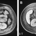 Imaging at Higher Magnetic Fields: 3 T Versus 1.5 T
 Magnetic Resonance Imaging of the Liver: Sequence Optimization and Artifacts
 Functional Magnetic Resonance Imaging of the Liver: Parametric Assessments Beyond Morphology
Stay updated, free articles. Join our Telegram channel

Full access? Get Clinical Tree


