15 Magnetic resonance imaging (MRI)
Definition of magnetic resonance imaging
| A non-invasive technique that uses radio-frequency radiation in the presence of a powerful magnetic field to produce high-quality images of the body in any plane |
| Acquisitions (Number of Excitations, Signal Averages) | The gathering of enough information to spatially encode one complete data set |
| Dephasing | When the RF pulse is switched off, the spinning protons go out of phase resulting in a reduction in the received signal |
| Echo Time (TE) |
• Gives the quantity of dephasing that happens between the excitation pulse and the echo
• The shorter the echo time, the lower the dephasing, the higher the proton density (T1) and the better the image
|
| Flip Angle (α) | The degree by which the proton is tipped in relation to the main magnetic field when a RF pulse is applied to it |
| Fourier Transform |
• A method of mathematically changing data, e.g. changing image space to k-space
|
| Gradient Echo |
• A basic pulse sequence that only uses magnetic field gradient reversal to re-phase the transverse magnetisation and produce echoes of the magnetic resonance signal
• Allows shorter repetition times and faster scanning
• Uses flip angles between 0° and 90°
|
| Image Space | An MRI image |
| Inversion Recovery (IR) |
• A basic pulse sequence of 180°, 90° and 180° which inverts the magnetisation and measures the time taken for the nuclei to return to the equilibrium
• The rate of recovery depends on the relaxation time T1
|
| k-space |
• The Fourier transform of an MRI image
• Gives the frequency and the phase encoding directions
|
| Larmor Equation | At a given field strength, the nuclei of different elements will precess at different frequencies, the equation is used to calculate the frequency of the RF pulse |
| Larmor Frequency | The rate at which the protons spin when a magnetic field is applied |
| Magnetic Field Gradient | The loss or increase of magnetic strength over distance controlled by the electrical current passing through the coil |
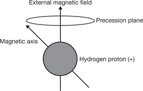
NoiseUnwanted electrical signals causing grain on the imagePrecessionIs the circular movement of the magnetic axis of a spinning proton which is prescribed when an external magnetic field is applied to the proton
Pulse SequenceThe bursts of electromagnetic energy produced by the radio-frequency coils


Radio-frequency (RF) PulseA burst of electromagnetic energy at right angles to the magnetic fieldRelaxation TimeThe time taken for the spinning protons to release the energy obtained and return to their original stateRepetition Time (TR)The time between the beginning of one radio-frequency pulse sequence to the start of the next, e.g. 300 ms or 500 ms at 1.5 TeslaResonanceWhen an object (a proton) responds to an alternating force (a radio-frequency signal) causing movementSaturated Recovery (SR)
SaturationThe maximum degree of magnetisation that can be achieved in a substanceSignal to Noise RatioImage quality = Signal (information required from image)/Noise (unwanted information on an image)Can be improved by:
Spatial EncodingThe prediction of the strength of the magnetic field and the movement of the protons at a set point along a gradientSpin Echo (SE)
Spin Polarisation
T1 Relaxation Time
T2 Relaxation Time
Tesla
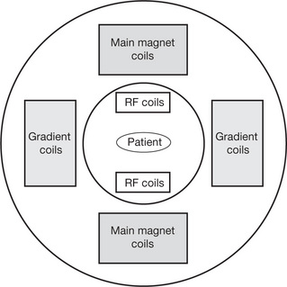
Fig. 15.2 MRI coil position (diagrammatic).
Related posts:

Stay updated, free articles. Join our Telegram channel

Full access? Get Clinical Tree


