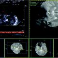
Fig. 7.1
Contrast-enhanced brain CT of a young patient with cerebral vein thrombosis, involving the left TS, the confluens of sinuses, and the distal end of the SSS. In the picture (a) is well evident the lack of endoluminal filling at the level of confluens of sinuses and left TS; in the picture (b) it can be seen the involvement of the posterior end of the SSS

Fig. 7.2
a–c TCCS from temporal bone window in axial access (same subject of the previous figure). There are two of the indirect signs suggestive for the diagnosis of cerebral venous thrombosis, i.e., the presence of venous collateral vessels behind the diencephalon in the picture (a) and the inverted flow direction of a vein, i.e., Right BVR in the post-peduncular segment with flow direction to the probe, opposite to the direction of the P3 PCA simultaneously sampled (b). Right SPS with flow direction to the probe (c)
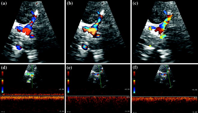
Fig. 7.3
a–e TCCS from the temporal bone window in axial scan (same subject of the previous figures). A further indirect sign of cerebral venous thrombosis can be seen, i.e., the increased flow velocity of a vein acting as alternative drainage route, as the SPS in this case. In the pictures (a) and (b), the medial end of the left SPS is insonated in Power mode with direction coding; in the picture (c), the same vein is insonated in Color mode with evident aliasing. In the pictures (d–e), the Doppler waveform is sampled in three sequential points of the medial segment of SPS, showing, particularly in picture (e), a markedly increased flow velocity with dispersion of frequencies in the waveform and orthograde direction
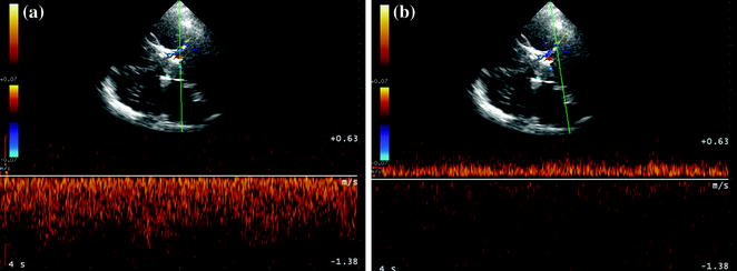
Fig. 7.4
a and b TCCS from temporal bone window in axial scan (same subjects of the previous figures). It shows another indirect sign of cerebral venous thrombosis, i.e., the inverted flow direction in a patent vein acting as alternative drainage route, in this example the right SPS, insonated in its medial end, which seems orthograde in the pre-terminal segment (a) and inverted in the previous segment (b)
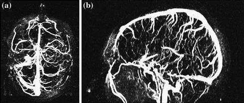
Fig. 7.5
Venous MRA with bidimensional reconstruction in a patient with cerebral venous thrombosis of the SRS, most of the GV and left TS
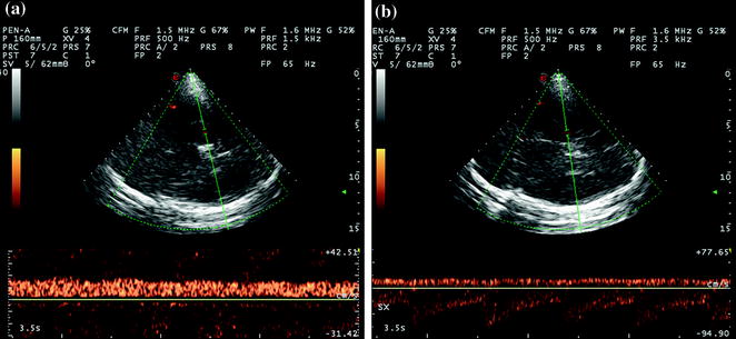
Fig. 7.6
a and b TCCS from the temporal bone window with insonation plane at the passage between mesencephalon and diencephalon in the same subject of the previous figure. It can be seen the post-peduncular segment of the BVR of both sides [the right BVR in the picture (a) and the left one in (b)] with inverted flow direction, congruent with the thrombotic occlusion of the GV-SRS
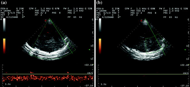
Fig. 7.7
a and b TCCS from temporal bone window in axial scan with midbrain insonation plane, oriented to the posterior cranial fossa (same subject of the previous two figures). Picture (a) shows the right TS, patent, and without abnormalities, (b) shows the site of left TS, lacking both for Doppler and for power-mode signals
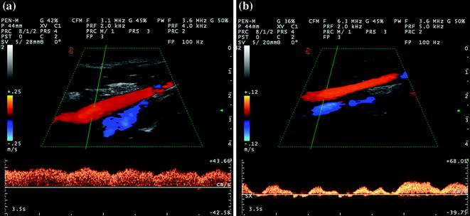
Fig. 7.8
Longitudinal scan of J3 IJV (same patients of the previous three figures). In picture (a), there is the right J3 IJV with a well-modulated Doppler waveform lacking abnormalities; in picture (b), there is the left J3 IJV, whose Doppler waveform is characterized by an alternating flow with superimposed phasic flow variations due to the breath cycle, both for the flow direction and for the velocity. As demonstrated in the picture, indirect signs can be seen at the level of the extracranial venous district, suggesting an intracranial site of disease, although these ones cannot be independently significant because of the wide variability of cerebral venous hemodynamics
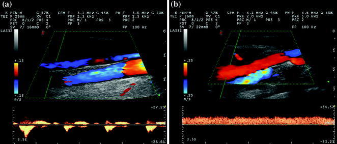
Fig. 7.9
Longitudinal scan of J2 IJV in Color mode in a patient with cerebral vein thrombosis involving the left TS. In picture (a), there is the left IJV with the corresponding Doppler waveform at alternating direction, phasic with the breath cycle (see also movies 7.6), while in picture (b), there is the right IJV, whose flow pattern is orthograde during the breath and heart cycles (see Movie 7.7)
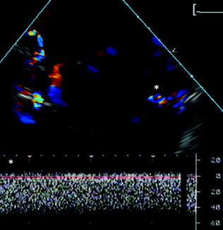
Fig. 7.10
TCCS from the right temporal bone window in axial scan, mesencephalic plane with posterior tilting of the probe, Color mode. The imaged patient has a severe TBI and a wide contusive injury with hemorrhagic evolution in the right brain hemisphere. It is possible to identify in Color mode the posterior end of the SRS with evident aliasing, as in the confluens of sinuses. The Doppler waveform corresponding to the SRS (below) shows an increased flow velocity and the loss of the regular pulsatility of the cerebral venous flow
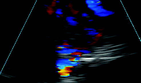
Fig. 7.11


TCCS from the right temporal bone window in axial scan, mesencephalic plane with posterior tilting of the probe, Color mode (same patient of the previous figure). Magnification of the confluens of sinuses and origin of the ipsilateral and contralateral TS. It is evident the aliasing effect on the contralateral TS compared with the ipsilateral one, as in the pre-terminal segment of the SRS
Related posts:
Stay updated, free articles. Join our Telegram channel
Full access? Get Clinical Tree
