Chapter 11. Male Pelvis
Patient Preparation
• Full bladder preparation for the urinary bladder or transabdominal prostate examination.
• Bowel preparation for the transrectal prostate examination.
• No preparation for the scrotal/testicular examination.
Equipment and Technical Factors
A high-frequency linear array is used for imaging the scrotum. Endorectal prostate imaging requires the use of the specialized transrectal transducer. The transrectal transducer should be adequately sheathed for the examination and appropriately disinfected after each use. Published guidelines for disinfection of endocavitary transducers are available. In addition, manufacturer specifications should be followed to avoid damaging the transducer.
Imaging Protocol
• Evaluate and document the longitudinal and transverse axes of the testicle regardless of the position of the testicle within the scrotum.
• Doppler (color, spectral) evaluation and comparison of blood flow in the testes should be documented. Low-flow settings should be used to avoid false-negative findings in cases of suspected torsion. Split-screen method allows flow in both testicles to be compared side by side. Doppler settings are the same for both testicles.
• Evaluate and document the prostate in the transverse axis from the superior aspect (base) to the inferior aspect (apex) and in the longitudinal axis from the midline to each lateral border.
• Longitudinal and transverse axes images of the bladder to demonstrate the overall size of the prostate may be done.
Sonographic Measurements
Testicle
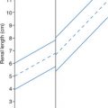
• Length: 4.0−5.0 cm
• Width: 2.0−3.0 cm
• Thickness (AP): 2.0−3.0 cm
Prostate
• Length: 2.0−4.0 cm
• Width: 3.9−5.3 cm
• Thickness: 2.1−3.4 cm
• Volume: 13.7 mL
• V = (π/6) × (L × W × T)
Seminal vesicles
• Length: 2.0−4.0 cm
• Diameter: 1.0 cm
| Male Pelvis | |||
|---|---|---|---|
| Sonographic Finding(s) | Clinical Presentation | Differential Diagnosis | Next Step |
Hypoechoic or heterogenous enlarged testicle Possible hydrocele noted Possible that testicular echogenicity and texture are normal Peripheral hyperemia may be noted No or decreased internal testicular flow demonstrated on color Doppler imaging | Scrotal swelling with pain | Testicular torsion | Compare affected testicle appearance with contralateral side Ensure Doppler device is set to normal testicle and not changed for affected testicle Because of peripheral hyperemia, flow may be detected in affected testicle but torsion should not be excluded because of this finding Testicle may demonstrate deceased flow from partial torsion or untwisting at time of exam |
| Cystic structure noted in epididymis, superior to testicle | Asymptomatic | Spermatocele Epididymal cyst | Evaluate for flow in cystic structure to ensure that the cyst is not a dilated vessel |
Fluid surrounds one or both testicles Amount of fluid may be small to massive | Asymptomatic Painful if related to rupture Possible scrotal enlargement if large amount of fluid
Related posts:Stay updated, free articles. Join our Telegram channel
Full access? Get Clinical Tree


| ||