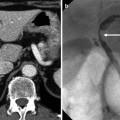
Fig. 25.1
Splenic angiosarcoma with spontaneous rupture in a 41-year-old male. (a) Noncontrast transverse CT image shows acute hematoma (short arrow) in the perisplenic space and hemoperitoneum in the perihepatic space (long arrow). A large mass lesion is also noted in the spleen. The portion of high attenuation (arrowheads) in the tumor suggests intratumoral hemorrhage. (b) Arterial (left) and portal (right) phase transverse CT images show peripheral enhancement of the mass (arrowheads)
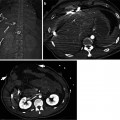
25.5.2 Splenic Angiosarcoma Involving Whole Spleen

Fig. 25.2
Splenic angiosarcoma in a 33-year-old female. Dynamic contrast-enhanced transverse CT images demonstrate a heterogeneously enhancing mass-like lesion replacing nearly whole portion of the enlarged spleen
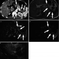
25.5.3 Splenic Angiosarcoma: MR Finding
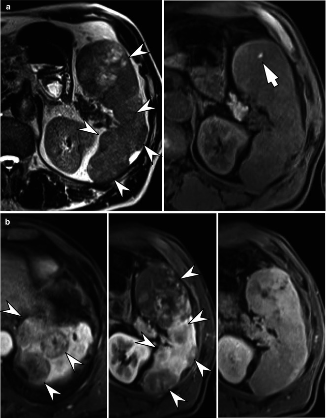
Fig. 25.3
Splenic angiosarcoma in a 63-year-old male. (a) T2-weighted transverse MRI (left) shows multiple masses (arrowheads) of variable signal intensities in the spleen. A bright dot (arrow) on fat-saturated T1-weighted transverse MRI (right) suggests intratumoral hemorrhage. (b) Contrast-enhanced portal phase T1-weighted transverse MRI (left and middle) clearly depict multiple splenic masses (arrowheads). Delayed phase (right) T1-weighted transverse MRI taken at 20 min after contrast media injection shows a delayed fill-in enhancement pattern of the masses
25.5.4 Splenic Lymphoma with Diffuse Involvement
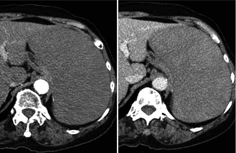
Fig. 25.4
A 72-year-old female with diffuse large B-cell lymphoma. Splenomegaly with diffuse low attenuation is noted both on arterial (left) and portal (right) phase CT images. There is no discernible focal lesion in the spleen
25.5.5 Splenic Lymphoma Appeared as a Focal Mass
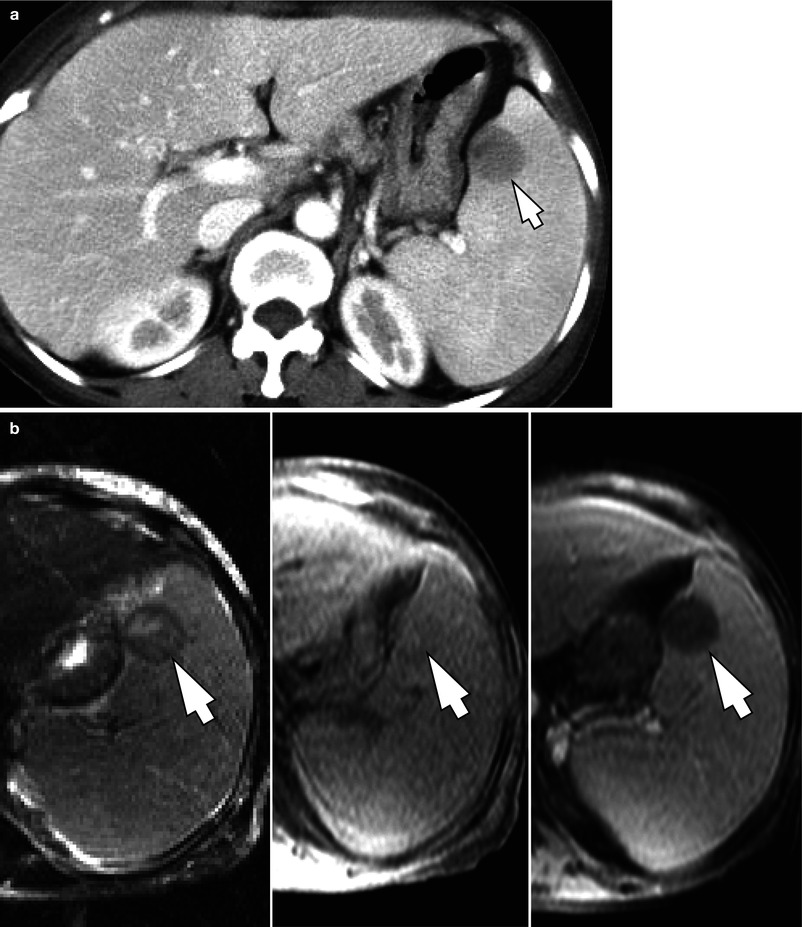
Fig. 25.5
A 77-year-old female with diffuse large B-cell lymphoma. (a) Contrast-enhanced transverse CT image demonstrates a 2.5 cm hypoattenuating mass (arrow) in the spleen. (b) The lesion (arrows) shows heterogeneous high signal intensity on T2-weighted transverse MRI (left), slightly low signal intensity on fat-saturated T1-weighted transverse MRI (middle), and poor enhancement after gadolinium injection (right


Related posts:


Stay updated, free articles. Join our Telegram channel
Full access? Get Clinical Tree
