Malrotation
Michael P. Federle, MD, FACR
Key Facts
Terminology
Rotational abnormality of the gut due to arrest of gut rotation and fixation during fetal development
Imaging
Many variations and classifications
Complete nonrotation
Jejunum to right of spine
Ileum to left of spine or in pelvis
Upper GI series with small bowel follow through
Normal barium enema does not exclude malrotation nor volvulus
Classic malrotation
Cecum lies in midline or to left
Fixed in position by bands from undersurface of liver (Ladd bands)
Ladd bands cross duodenum and may obstruct it
Oral contrast medium ends abruptly or in “corkscrew” pattern within duodenum
CT: Superior mesenteric vein (SMV) ventral to or on left of SMA
Volvulus of midgut indicated by “whirlpool” or “swirl” sign of twisted mesenteric vessels and bowel
CT shows congenital anomalies and complications
Top Differential Diagnoses
Paraduodenal hernia
Clinical Issues
Up to 40% diagnosed by 1 week of age
50% by 1 month
75% by 1 year
25% after 1 year
Some cases of volvulus or obstruction occur in older children or adults
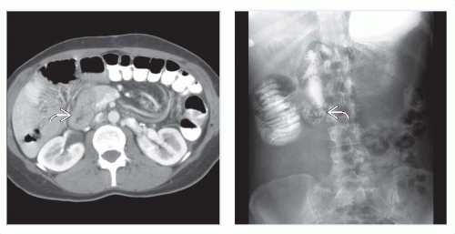 (Left) 27-year-old man with pain and vomiting. Axial CECT shows all of the small bowel, even the duodenum  , lying to the right of midline. The cecum is midline, and the remaining colon lies to the left of midline. (Right) Upper GI series in the same patient shows partial obstruction at the level of the distal duodenum. The duodenum never crosses the midline but has a peculiar “Z” or “corkscrew” configuration , lying to the right of midline. The cecum is midline, and the remaining colon lies to the left of midline. (Right) Upper GI series in the same patient shows partial obstruction at the level of the distal duodenum. The duodenum never crosses the midline but has a peculiar “Z” or “corkscrew” configuration  . Malrotation and duodenal obstruction by bands were confirmed at surgery. . Malrotation and duodenal obstruction by bands were confirmed at surgery. |
 (Left)
Get Clinical Tree app for offline access
Related posts:Stay updated, free articles. Join our Telegram channel
Full access? Get Clinical Tree


|