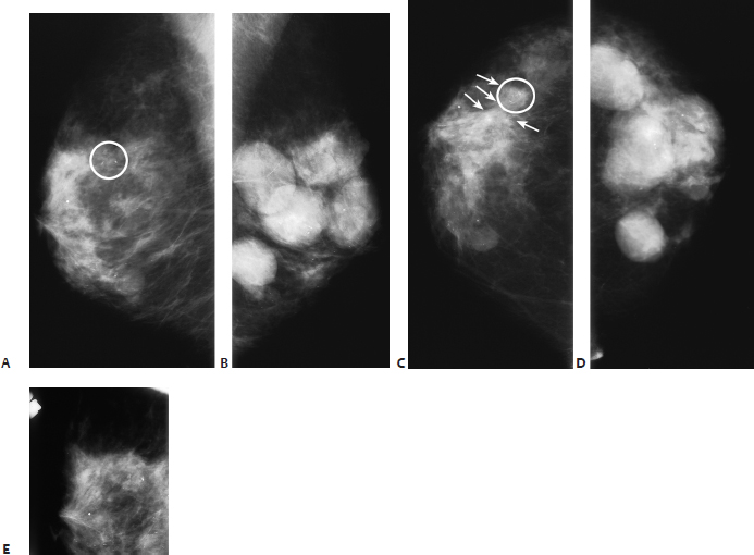
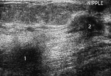
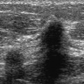
31 Mammogram Underestimates Tumor Size A 69-year-old woman presents for screening mammogram. • Bilateral breast: multiple lumps Calcifications (Fig. 31.1) • Type: pleomorphic/heterogeneous • Distribution: grouped/clustered Fig. 31.1 The right breast has a cluster of heterogeneous calcifications (circle) in the upper outer breast. These calcifications are associated with architectural distortion in the CC view. The left breast has multiple round and oval masses that have been stable for several years. (A) Right MLO mammogram. (B) Left MLO mammogram. (C) Right CC mammogram. (D) Left CC mammogram. (E) Right ML spot magnification mammogram. Frequency • 12 MHz Mass (Fig. 31.2) • Margin: ill defined • Echogenicity: hypoechoic • Retrotumoral acoustic appearance: posterior shadowing distal to mass • Shape: irregular Fig. 31.2 Right radial breast sonogram. Two hypoechoic irregular masses are present. Mass 1 is in the upper outer quadrant, and mass 2 is next to the nipple. • Mixed ductal and lobular carcinoma • Two separate foci of mixed ductal and lobular carcinoma • BI-RADS assessment category 5, highly suggestive of malignancy • In this case, it was not clear if the architectural distortion was associated with the abnormal calcifications, as the distortion was visible in only one view. Furthermore, the architectural distortion appeared much larger than the area of the calcifications. Sonography may be helpful when there is a mammographic imaging discrepancy. In this case, the abnormal mammographic calcifications and architectural distortion correspond to multifocal disease. Baker JA, Soo MS. The evolving role of sonography in evaluating solid breast masses. Semin Ultrasound CT MR 2000;21:286–296 Buchberger W, Niehoff A, Obrist P, DeKoekkoek-Doll P, Dünser M. Clinically and mammographically occult breast lesions: detection and classification with high-resolution sonography. Semin Ultrasound CT MR 2000;21:325–336 A 45-year-old woman presents with thickening in the right breast, which she thinks might be a pulled muscle. • Right breast: large, mildly tender firm area extending from the 9 o’clock to the 12 o’clock positions in the right breast • Left breast: normal exam Mass (Fig. 31.3) • Margin: ill defined • Shape: oval • Density: isodense
Case 31.1: Mammogram Underestimates Tumor Size
Case History
Physical Examination
Mammogram
Ultrasound
Pathology
Management
Pearls and Pitfalls
Suggested Reading
Case 31.2: Mammogram Underestimates Tumor Size
Case History
Physical Examination
Mammogram
Related posts:


Stay updated, free articles. Join our Telegram channel

Full access? Get Clinical Tree


