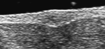
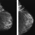
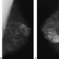
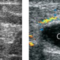
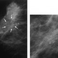
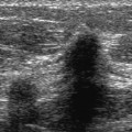
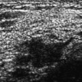
32 Mass in Unusual Locations A 42-year-old woman presents with a small, superficial left breast lump. • Left breast: a soft, superficial lump at the 12 o’clock position • Right breast: normal exam Frequency • 10 MHz Mass (Fig. 32.1) • Margin: ill defined • Echogenicity: heterogeneous • Retrotumoral acoustic appearance: posterior shadowing distal to mass • Shape: ellipsoid Fig. 32.1 Left antiradial breast sonogram. The palpable lump corresponds to an ill-defined oval mass with heterogeneous echogenicity that is within the skin. • Smooth muscle hamartoma • BI-RADS assessment category 3, probably benign; short-interval follow-up
Case 32.1: Mass in Unusual Locations
Case History
Physical Examination
Ultrasound
Pathology
Management
Radiology Key
Fastest Radiology Insight Engine

