Fig. 1
Cutting apparatus used for longitudinal sectioning of hair samples. (a) Image of the in-house built cutting apparatus and (b) schematic of the cutting apparatus
Following decontamination, place a single hair sample into one of the grooves of the cutting block and fix at one end with a small piece of double-sided tape, as shown in Fig. 2a (see Notes 3 – 5 ).

Fig. 2
Method for preparing longitudinal sections of single hair samples. (a) Hair sample attached to the cutting block with double-sided tape , (b) preparing longitudinal sections of single hair samples using in-house built cutting apparatus, (c) longitudinal sections mounted on conductive glass slide with double-sided copper tape
While holding the other end with a gloved finger, slowly run the cutting device along the length of the hair (Fig. 2b).
Place another piece of double-sided tape on the other end of the hair and transfer onto double-sided copper tape mounted on a ITO glass slide, press the hair into the tape using a clean glass slide (Fig. 2c).
Use a Leica DMRX microscope equipped with a digital camera to determine if the hair section has been successfully transferred onto the glass slide.
3.3 Preparation of Cross-Sections
- 1.
Following decontamination, thread the hair sample through the lid of the tube and using a paper clip, attach two small magnets at the end of the single hair sample. Attach two more magnets at the opposite end of the hair sample.
- 2.
Place the hair sample into a plastic tube that contains a 10% solution (w/v) of gelatin, as shown in Fig. 3b (see Note 6 ).

Fig. 3
Method for embedding single hair samples. (a) Materials required for the embedding of single intact hair samples, (b) single hair sample clipped between magnets and placed in a plastic tube filled with embedding material, (c) embedded hair sample following snap freezing, and (d) cryo-sectioning of embedded hair samples
- 3.
Snap-freeze the contents by placing the plastic tube in liquid nitrogen for 30 s (Fig. 3c).
- 4.
Remove the embedded hair from the plastic tube and cut into 1 cm blocks (the average growth of hair being 1 cm/month), mount one of the blocks onto a cryo-microtome stage using a drop of water.
- 5.
Section the embedded hair samples at −20 °C using a cryo-microtome to produce 12 μm thick sections, thaw mount sections onto a clean indium tin oxide (ITO) glass slide (see Note 7 ).
- 6.
Inspect the sections using a Leica DMRX microscope equipped with a digital camera to determine the position of the hair cross-section and mark the location on the opposite side of the glass slide using a permanent marker pen.
3.4 Matrix Deposition for MALDI-MS/MS Imaging
- 1.
Place the sample in the ImagePrep matrix application device.
- 2.
Fill the bottle with the matrix solution.
- 3.
Select the manufacturer’s method for spraying CHCA and start the sequence.
- 4.
Use a Leica DMRX microscope equipped with a digital camera to inspect the crystal coverage.
3.5 Metal Deposition for MetA-SIMS Imaging
- 1.
Place the sample in the chamber of the Quorum Technologies sputter coater and close the lid.
- 2.
Select the density of the metal (19.30 g/cm3 in the case of gold) and the desired thickness (1 nm) on the film thickness monitor.
- 3.
Set the discharge voltage to 1.5 kV and the plasma current to 25 mA in order to achieve a homogenous coating.
- 4.
Start the sequence (operate in automatic mode), once the sequence is complete vent the instrument and remove the sample (see Note 8 ).
3.6 MALDI-MS/MS Imaging
- 1.
Calibrate the instrument prior to analysis with either a standard mixture of polyethylene glycol (PEG 200-3000) in water mixed with matrix or a saturated solution of red phosphorus in acetone.
- 2.
Spot 0.5 μL of a cocaine base standard (100 ng/μL in 70% MeOH) onto a MALDI target plate, followed by 0.5 μL of the matrix solution.
- 3.
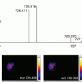
Optimize the instrumental settings using the cocaine base standard such as the laser power and collision energy (trap cell). The optimal settings were as follows: laser power 250 (200 Hz) and collision energy 10 eV (monitor the main product ion of cocaine at m/z 182 formed by neutral loss of benzoic acid).
- 4.
Following method optimization , attach the sample onto a glass slide adaptor using double-sided tape and scan using a flatbed scanner to produce a digital image, ensure the image quality is around 600 dpi or better.
- 5.
Import the digital image of the sample into the MALDI imaging pattern creator software, to define the area to be imaged and the spatial resolution (150 × 50 μm).
- 6.
Set up the MS/MS imaging method in the MassLynx 4.1 software. The settings were as follows: positive ion mode, V-mode, mass range m/z 50–350, also use the previously optimized settings (see step 3).Related posts:
 “A Future Amalgamation Between the Scientist and the Clinician?”
 Peptide Imaging: Maximizing Peptide Yield, Optimization of the “Peptide Mass Fingerprint”
 MALDI-MS Imaging in the Study of Glomerulonephritis
 DESI Mass Spectrometry Imaging (MSI)
 Imaging MS of Rodent Ocular Tissues and the Optic Nerve
 Laser Ablation Inductively Coupled Plasma Mass Spectrometry Imaging of Plant Metabolites

Stay updated, free articles. Join our Telegram channel

Full access? Get Clinical Tree


