Mastoiditis
Scott S. Abedi
CLINICAL HISTORY
6-year-old girl with spiking fevers, left ear swelling, and bilateral purulent ear drainage despite antibiotics and bilateral pressure equalization tube placement.
FINDINGS
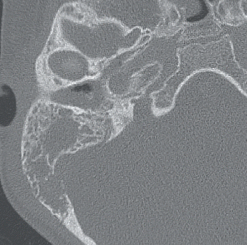
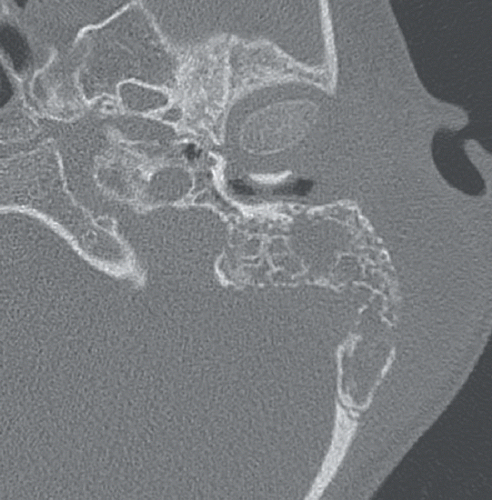
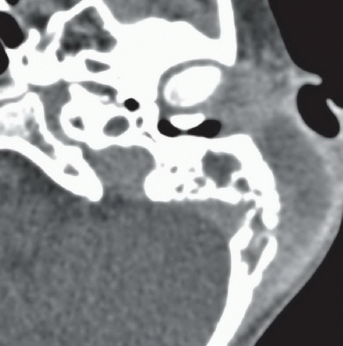
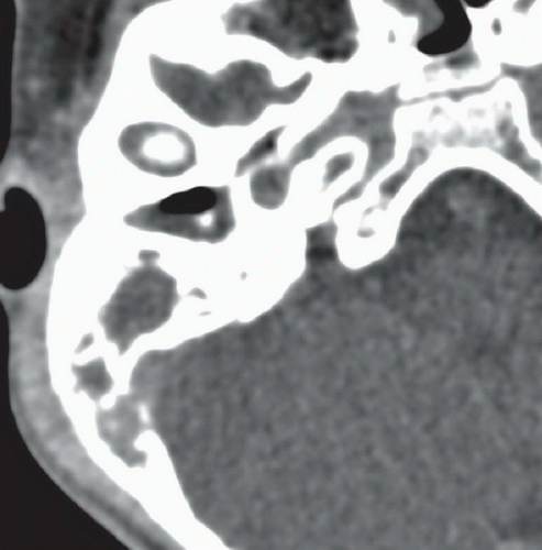
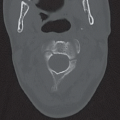
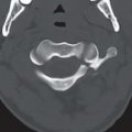
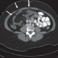
Figures 5A and 5B: Axial contrast-enhanced CT through the temporal bones displayed in bone windows. There are diffuse permeative osseous changes through the bilateral mastoid air cells as well as complete opacification of both the mastoids and the middle ears. Notice the asymmetric fullness along the lateral margin of the left mastoid. Figures 5C and 5D: Soft tissue windowing at these same levels clearly depicts a peripherally enhancing subperiosteal fluid collection along the lateral margin of the left mastoids.
Stay updated, free articles. Join our Telegram channel

Full access? Get Clinical Tree


