Mediastinal Hematoma
Katherine R. Birchard
CLINICAL HISTORY
36-year-old male after a motor vehicle accident.
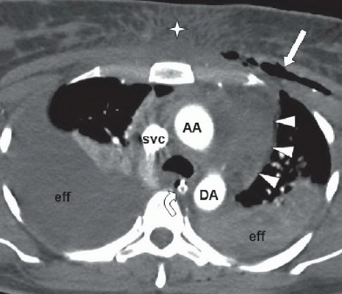 FIGURE 42A |
FINDINGS
Figure 42A: Axial contrast-enhanced CT image of the chest just below the aortic arch shows mediastinal hematoma (arrowheads). Ascending aorta (AA), descending aorta (DA), and superior vena cava (SVC) are intact. Large right pleural effusion is present, and higher density left effusion is likely hemothorax (eff). Note anterior chest wall contusion (star), subcutaneous air (arrow), and nasogastric tube (curved arrow).
DIFFERENTIAL DIAGNOSIS
Mediastinal hematoma, thymoma, lipoma.
DIAGNOSIS
Related posts:


Stay updated, free articles. Join our Telegram channel

Full access? Get Clinical Tree


