Mesenteric Injury
Ho Chia Ming
Ellie R. Lee
CLINICAL HISTORY
61-year-old man with severe abdominal trauma from a motor vehicle accident.
FINDINGS
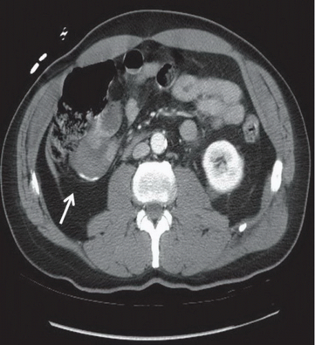
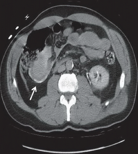
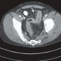
Figure 55A: Axial contrast-enhanced CT image performed in the portal venous phase demonstrates active extravasation of contrast from an ileocolic arterial branch into the mesentery, showing similar attenuation with the contrast within the vessel lumen. Mesenteric hematoma with high attenuation is identified (arrow). Figure 55B: Delayed axial contrast-enhanced CT image at the same level demonstrates increased density and size of the mesenteric hematoma, indicating acute mesenteric injury and active bleeding (arrow).
DIFFERENTIAL DIAGNOSIS
Mesenteric injury, bowel injury, renal or ureteral injury.

