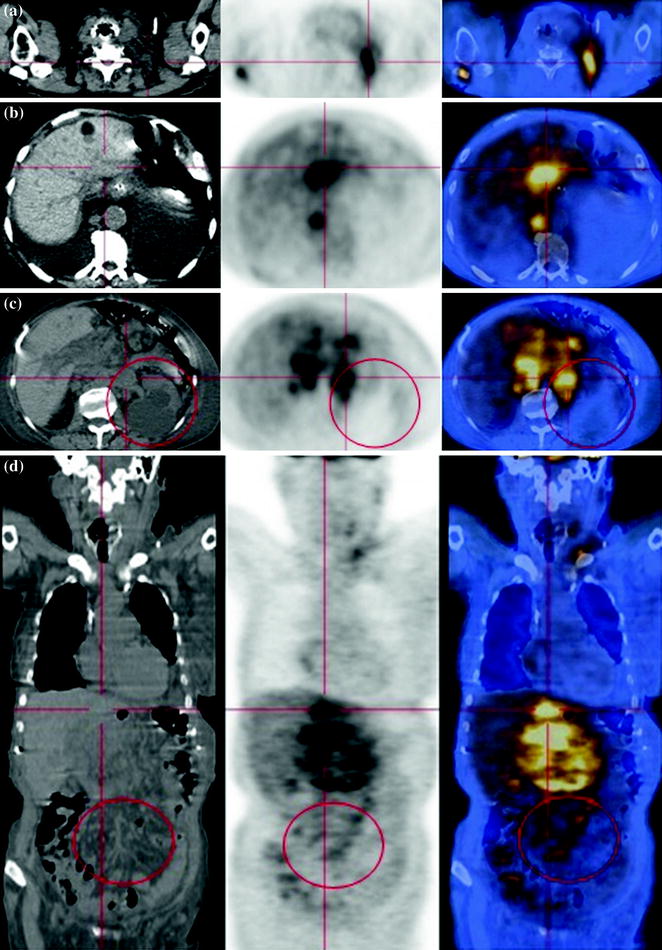
Fig. 76.1
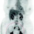
The PET scan shows multiple enlarged lymph nodes with high glucose metabolism in the celiac-mesenteric-aortic, left lumbo-aortic regions; bilaterally in the iliac region, even though more pronounced on the left side, SUVmax 14
Presence of numerous mediastinal anterior and posterior nodes with high metabolism, SUVmax 7.5; nodes can be found in the clavicular fossa and the latero-cervical region on both sides, with greater involvement on the left, SUVmax 7.
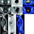
Multiple skeletal lesions with a high FDG uptake, with involvement of the right scapula, some dorso-lumbar vertebrae, femurs and pelvis, more pronounced in the sacrum and the sacroiliac articulation, SUVmax 10.
The left kidney is excluded for severe obstructive hydronephrosis due to the infiltration of the ureter. The right pielo-ureteral region shows a lesser dilatation that does not reflect obstructive component.
76.4 Conclusions
The PET scan shows a progression of disease with various skeletal metastasis and lymph node diffuse involvement, due to poor response to chemotherapy. See Figs. 76.2, 76.3, 76.4.
