
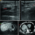
Fig. 18.1
Ultrasound-guided percutaneous microwave ablation (MWA) in a 55-year-old female patient with angiomyolipoma in the upper pole of the right kidney. (a ) The lesion is hyperechoic and with well-defined borders in ultrasound (arrow). (b) The lesion showed heterogeneous hypo-enhancement in the arterial phase of contrast-enhanced ultrasound and the dimension is 5.7 × 5.4 cm (arrows). (c) After ablation, the lesion showed no enhancement in the arterial phase with contrast-enhanced MRI in transversal scan (arrows). (d) After ablation, the lesion showed no enhancement in the delayed phase with contrast-enhanced MRI in coronal scan (arrows). The significance of 1 and 2 in the Fig. 18.1 (a) and (b) is the two perpendicular diameters
Table 18.1
Basic characteristics and clinical results of patients with renal AMLs treated with ablation
Author | No. of Pts (nodules) | Size (cm) | Treatment type | Guided methods | Follow-up (months) | Results (no. of recurrence/persistence) | Major complications (patients) |
|---|---|---|---|---|---|---|---|
Sooriakumaran et al. [26] | 4 (4) | 9.5 (9–19) | RFA | N/A | 9 (2–13) | 0/4 | 0 |
Gregory et al. [27] | 4 (4) | 15.1 (6.1–32.4) | RFA | CT | 48 | 0/4 | 0 |
Prevoo et al. [28] | 1 (1) | 4.5 | RFA | CT | 12 | 1/0 | 0 |
Castle et al. [29] | 15 (15) | 2.6 (1.0–3.7) | RFA | Laparoscopic (5) or CT (10) | 21.1 (1.5–72) | 15/0 | 2 |
Byrd et al. [30] | 6 (12) | 4.2 (2.5–7.0) | Cryoablation | Laparoscopic | N/A | 7/0 | 0 |
Johnson et al. [13] | 3 (3) | (1.2–2.5) | Cryoablation | CT | 5–36 | 2/1 | 0 |
Han et al. [35] | 14 (19) | 3.4 (0.8–6.1) | MWA | US | 10 (6–36) | 15/4 | 2 |
18.6 Conclusion
Because of the nonaggressive biologic behavior of these benign renal tumors, there is increasing interest in minimally invasive treatment modalities, particularly for the elderly, the infirm, and patients with comorbid conditions. MWA may be an alternative minimally invasive technique for the treatment of renal AML. However, the studies of large sample and long-term follow-up period are necessary to determine efficacy, and safety.
References
1.
2.
3.
4.
Steiner MS, Goldman SM, Fishman EK, Marshall FF. The natural history of renal angiomyolipoma. J Urol. 1993;150:1782–6.PubMed
Related posts:
 Microwave Ablation Combined with Cellular Immunotherapy for Hepatocellular Carcinoma
 Microwave Ablation for Malignant Liver Tumors Adjacent to the Hepatic Hilum
 Ultrasound-Guided Microwave Ablation for Superficial Malignant Tumors
 Microwave Ablation for Adenomyosis
 Clinical Application of Three-Dimensional Visualization Techniques in Microwave Ablation for Liver Cancer
 Microwave Ablation in Renal Cell Carcinoma
Stay updated, free articles. Join our Telegram channel

Full access? Get Clinical Tree


