Miscellaneous Bone Lesions
Clyde A. Helms
There are a host of bony conditions, diseases, and syndromes that do not fit conveniently into any of the preceding chapters, yet should be given some mention in an attempted overview of musculoskeletal radiology. These are listed alphabetically for lack of a more scientific basis.
Achondroplasia
The most common cause of dwarfism is achondroplasia, a congenital, hereditary disease of failure of endochondral bone formation. The femurs and humeri are more profoundly affected than the other long bones, although the entire skeleton is abnormal. A characteristic finding is that the spine typically has narrowing of the interpedicular distances in a caudal direction (Fig. 46.1), the opposite of normal, in which the interpedicular distances get progressively wider as one proceeds down the spine. The long bones are short but have normal width, giving them a thick appearance.
Avascular Necrosis (Osteonecrosis)
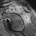
The term “avascular necrosis” (AVN) or osteonecrosis refers to the lack of blood supply with subsequent bone death and ensuing bony collapse in an articular surface. The etiology of AVN is an extensive differential that most commonly includes trauma, steroids, aspirin, renal disease, collagen vascular diseases, alcoholism, and idiopathic causes (Table 46.1) (1). The radiographic appearance ranges from patchy sclerosis (Fig. 46.2A) to articular surface collapse and fragmentation (Fig. 46.3). Just before collapse, a subchondral lucency is occasionally seen (Fig. 46.4); however, this is a late and inconstant sign of AVN. MR is extremely valuable in demonstrating the presence and extent of AVN (Fig. 46.2B), even when plain films are apparently normal. MR is currently considered to be the most efficacious way to evaluate a joint for AVN (2). It is useful not only in AVN of the hip but also in the knee, wrist, foot, and ankle.
Hypertrophic Pulmonary Osteoarthropathy
Hypertrophic pulmonary osteoarthropathy is manifested by clubbing of the fingers and periostitis, usually in the upper and lower extremities (Fig. 46.5), which might or might not be associated with bone pain. It is most commonly seen in patients with lung cancer, but many other etiologies have been reported, including bronchiectasis, GI disorders, and liver disease. The actual mechanism of formation of periostitis secondary to a distant malignancy or other process is unknown. The differential diagnosis for periostitis in a long bone without an underlying bony abnormality would include hypertrophic pulmonary osteoarthropathy, venous stasis, thyroid acropachy, pachydermoperiostosis, and trauma (Table 46.2).
Melorheostosis
Melorheostosis is a rare, idiopathic disorder characterized by thickened cortical new bone that accumulates near the ends of long bones, usually only on one side of the bone, and has an appearance likened to “dripping candle wax” (Fig. 46.6). It can affect several adjacent bones and can be symptomatic.
Mucopolysaccharidoses (Morquio, Hurler, and Hunter Syndromes)
The mucopolysaccharidoses are a group of inherited diseases characterized by abnormal storage and excretion in the urine of various mucopolysaccharidoses such as keratin sulfate (Morquio) and heparan sulfate (Hurler). These patients have short stature, primarily from shortened spines, and characteristic plain film findings. In the spine, patients with Morquio have platyspondyly (generalized flattening of the vertebral bodies) with a central anterior projection or “beak” off the
vertebral body, as viewed on a lateral plain film (Fig. 46.7). Hurler and Hunter show platyspondyly with a beak that is anteroinferiorly positioned (Fig. 46.8). The pelvis in these disorders is similar in appearance to that of achondroplasts, with wide, flared iliac wings and broad femoral necks. A characteristic finding in the hands is a pointed proximal fifth metacarpal base that has a notched appearance to the ulnar aspect (Fig. 46.9).
vertebral body, as viewed on a lateral plain film (Fig. 46.7). Hurler and Hunter show platyspondyly with a beak that is anteroinferiorly positioned (Fig. 46.8). The pelvis in these disorders is similar in appearance to that of achondroplasts, with wide, flared iliac wings and broad femoral necks. A characteristic finding in the hands is a pointed proximal fifth metacarpal base that has a notched appearance to the ulnar aspect (Fig. 46.9).
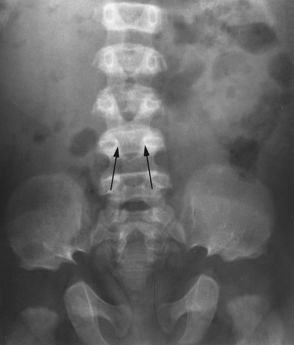 Figure 46.1. Achondroplasia. An anteroposterior plain film of the spine in this patient with achondroplasia demonstrates narrowing of the interpedicular distance (arrows) in a caudal direction, which is characteristic of this disorder. Ordinarily, the interpedicular distance widens in each vertebra in a caudal direction. |
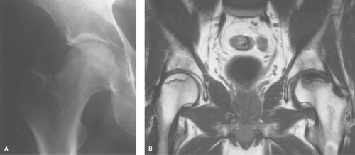 Figure 46.2. Avascular Necrosis (AVN). A. A plain film of the hip in this patient with AVN shows faint, patchy sclerosis throughout the femoral head. This is a relatively early plain film finding for AVN. B. Coronal T1-weighted (TR 500; TE 28) MR image shows typical findings in AVN. Diffuse low signal in the left hip is noted, which has more extensive involvement than the right. The right hip has a low-signal serpiginous rim which is characteristic of AVN. |
 Figure 46.3. Avascular Necrosis (AVN). An AP plain film of the shoulder reveals articular surface collapse in this patient who was treated with steroids for systemic lupus erythematosus. This is an advanced stage of AVN. |
Table 46.1 Common Causes of Avascular Necrosis | ||||||
|---|---|---|---|---|---|---|
|
Related posts:


Stay updated, free articles. Join our Telegram channel

Full access? Get Clinical Tree


