Fig. 1.1
Endocardial cushion embryological development of the atrioventricular valves occurs between gestational weeks 5 and 8 [Figures a–c]
The mitral valve apparatus is a complicated structure consisting of the leaflets, the annulus, the chordae tendineae, and the papillary muscles. Pathology of any of these aspects can result in mitral valve dysfunction. Chordae attach along the entire leaflet coaptation line and insert into the papillary muscle heads (Fig. 1.2). The primary or “marginal” chordae insert on the free margin of the valve leaflets. Rupture of these chordae always causes valve insufficiency, which is often severe. The secondary chordae (“strut chordae”) insert on the rugged ventricular surface of the leaflets. The secondary chordae may promote contraction of the ventricular longitudinal fibers, supporting the ventricular contraction. The tertiary chordae originate directly from the ventricular wall and insert only in the basal part of the posterior leaflet. The function of these chordae is unknown; they seem to reduce leaflet mobility by anchoring the leaflet to the ventricular wall.
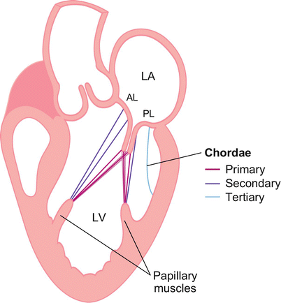
Fig. 1.2
Mitral subvalvular apparatus including chordal attachments
The papillary muscles, which are the muscular part of the mitral apparatus, originate from the distal one third of the ventricular wall. There are two dominant papillary muscles, anterolateral and posteromedial. These provide chordae to a portion of each leaflet. The posteromedial papillary muscle is perfused only from the right coronary artery, so it is more susceptible to ischemic insult than the anterolateral papillary muscle, which has a dual blood supply.
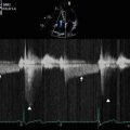
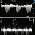
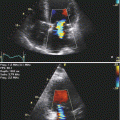
The mitral annulus is part of the fibrous skeleton of the heart. It is a complex, saddle-shaped, nonplanar geometric structure with greater commissure-commissural diameter (long axis) and smaller septolateral diameter (short axis) (Fig. 1.3). The leaflets do not normally protrude above the annular plane. Three-dimensional (3D) echocardiography has revolutionized visualization and appreciation of the mitral valve annulus (Fig. 1.4).
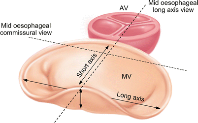
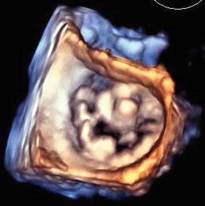
Fig. 1.3
Saddle-shaped structure of the mitral valve
Fig. 1.4
Three-dimensional (3D) transesophageal echo (TEE) image of the mitral valve
Related posts:

Stay updated, free articles. Join our Telegram channel

Full access? Get Clinical Tree


