Abstract
The term monochorionic refers to a multiple gestation with one placental disk (or chorion), and the term diamniotic describes the presence of two distinct amniotic cavities. By definition, monochorionic twin pregnancies are monozygotic. In the early first trimester, monochorionic diamniotic twin pregnancies can be identified by the presence of one gestational sac containing two fetuses. Monochorionic diamniotic pregnancies are characterized by the presence of two fetuses of the same gender, a single placenta, a thin two-layered intertwin membrane, and a characteristic T-shaped insertion of the intertwin membrane at the placental surface. In addition to risk of preterm delivery and growth disorders present in all twin gestations, monochorionic diamniotic pregnancies have many complications unique to the twinning process and monochorionic placentation, including an increased risk of structural anomalies, twin transfusion syndrome (TTS), and twin reversed arterial perfusion sequence. Serial prenatal ultrasound evaluations are essential in the management of monochorionic twin pregnancies.
Keywords
monochorionic, diamniotic, twin, multiple pregnancy
Introduction
Compared with dichorionic twin gestations, monochorionic twin gestations, whether diamniotic or monoamniotic, have an increased risk of adverse pregnancy outcomes. Ultrasound (US) plays a vital role in prenatal determination of chorionicity and in the management of monochorionic diamniotic twin pregnancies.
Disease
Definition
The term monochorionic refers to a multiple gestation with one placental disk (or chorion), and the term diamniotic describes the presence of two distinct amniotic cavities. In a monochorionic diamniotic twin pregnancy, there is one shared placenta, and each fetus has its own amniotic sac. By definition, monochorionic twin pregnancies are monozygotic.
Prevalence and Epidemiology
Twin gestations accounted for almost 3.4% of live births in the United States in 2014. The frequency of monozygotic twins is constant worldwide at 4 : 1000 births. Approximately two-thirds of monozygotic twin gestations have monochorionic placentation and diamniotic membrane composition. Dizygotic monochorionic diamniotic twin gestations resulting from assisted reproductive technology have been described in case reports.
Etiology and Pathophysiology
Monozygotic twinning results from the division of a single fertilized ovum into two distinct fetuses. Chorionicity and amnionicity of monozygotic gestations are determined by the time at which division of the fertilized ovum occurs. If twinning occurs during the first 2 to 3 days, it precedes the separation of cells that eventually become the chorion and results in a dichorionic diamniotic pregnancy. After approximately 3 days, twinning cannot split the chorionic cavity, and division of an ovum results in monochorionic placentation. If the split occurs between the third and eighth days, a monochorionic diamniotic pregnancy develops, whereas divisions that occur between the eighth and thirteenth days result in a monochorionic monoamniotic pregnancy.
Manifestations of Disease
Clinical Presentation
Multifetal gestations should be suspected if uterine size is greater than expected. Similarly, the possibility of a multiple gestation should be considered if elevated concentrations of maternal serum analytes, such as beta-human chorionic gonadotropin, alpha-fetoprotein, and other serum aneuploidy markers, are noted in the first or second trimester. When a multiple gestation is suspected clinically, US should be performed to assess plurality and, if a multiple gestation is confirmed, to determine chorionicity and amnionicity. There are no non-US clinical findings that can differentiate monochorionic from dichorionic twin gestations.
Imaging Technique and Findings
Ultrasound.
In the first trimester, monochorionic twin pregnancies can be easily detected on US. Between 6 and 10 weeks’ gestation, a monochorionic twin pregnancy can be identified by the presence of a single gestational sac containing two fetuses. The presence or absence of an intertwin membrane may be difficult to determine in the middle of the first trimester; however, the number of yolk sacs can also be used as an indirect method of determining amnionicity early in gestation. The presence of two yolk sacs is diagnostic of a diamniotic twin pregnancy, whereas a single yolk sac suggests a monoamniotic twin pregnancy.
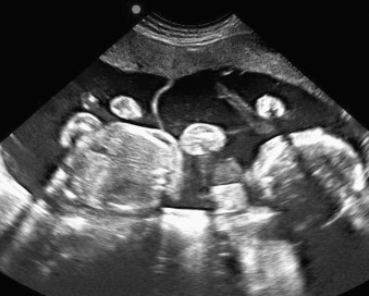
Later in gestation, monochorionic diamniotic pregnancies can be differentiated from dichorionic diamniotic and monochorionic monoamniotic twin pregnancies through a systematic evaluation of the placental number, fetal gender, and intertwin membrane. In monochorionic pregnancies, there is a single placenta and a thin intertwin membrane that inserts directly into the placenta; this finding, known as the “T” sign ( Figs. 160.1 and 160.2 ), has 100% sensitivity and greater than 98% specificity for detecting monochorionic diamniotic gestations. In contrast, the intertwin membrane of dichorionic gestations has a characteristic twin peak or “lambda” sign, and monoamniotic gestations lack an intertwin membrane altogether.

Although the findings of concordant fetal gender and a single placental mass support the diagnosis of monochorionic placentation, these findings are nonspecific and may also be detected in dizygotic and dichorionic twin pregnancies. In a study of more than 400 twin pregnancies undergoing US at less than 24 weeks’ gestation at a single tertiary care center, evaluation of a combination of features including placental number and location, membrane insertion (including twin peak and “T” signs), and fetal gender resulted in accurate prenatal diagnosis of chorionicity in greater than 95% of cases.
Careful US examination of the dividing membrane may also be used to aid in the diagnosis of chorionicity in midgestation. Specifically, the thickness and number of layers of the intertwin membrane can be assessed in a highly magnified image obtained with the membrane perpendicular to the US beam. If the membrane is thin and has only two layers, the pregnancy is monochorionic; in contrast, the intertwin membrane of monochorionic twin pregnancies is thicker (greater than 2 mm) or has three to four visible layers. Determination of the number of membrane layers has been reported to have greater than 98% accuracy in prenatal determination of chorionicity.
When attempting to determine amnionicity of a monochorionic gestation, the thin dividing membranes of some monochorionic diamniotic pregnancies may not be visualized on US because severe oligohydramnios causes them to be closely apposed to the fetus in that sac. A “stuck twin” appearance results, with the trapped fetus remaining firmly held against the uterine wall despite changes in maternal position. Diagnosis of this condition confirms the presence of a diamniotic gestation, which should be distinguished from a monoamniotic gestation with an absent dividing membrane. In the latter situation, free movement of both twins and entanglement of their umbilical cords can be identified.
Differential Diagnosis From Imaging Findings
The differential diagnosis of monochorionic diamniotic twin gestations includes twin gestations with alternative placentation and membrane composition. Specifically, monochorionic diamniotic pregnancies can be differentiated from dichorionic diamniotic pregnancies by assessing fetal gender, placental number, and characteristics of the intertwin membrane as described earlier. By definition, twins of a monochorionic pregnancy are of the same gender and share a single placenta (see Figs. 160.1 and 160.2 ). The intertwin membrane of monochorionic diamniotic pregnancies is composed of only two layers and appears thinner than the dividing membrane of dichorionic pregnancies. The placental insertion of a monochorionic membrane has the characteristic appearance of a “T” sign (see Figs. 160.1 and 160.2 ). In contrast, fetuses of a dichorionic twin pregnancy each have a separate placental mass, may be of different gender, and are separated by a thick four-layer membrane with a characteristic twin peak or lambda sign at its placental insertion.
Related posts:


Stay updated, free articles. Join our Telegram channel

Full access? Get Clinical Tree


