

The articles in this first issue—all written by international experts in the field—focus on topics directly relevant to patient care, thereby emphasizing once again the rapidly growing clinical importance of neonatal MRI. This issue is divided into three logical sections that cover all relevant anatomical and functional regions of the neonate. The first part covers, in accord with clinical appearance, the majority of MRI exams indicated for injuries and disorders of the brain and spine: hypoxic ischemic brain injury in preterm and term infants is discussed, as well as seizures, birth-related and inflicted head injuries, head trauma, and metabolic diseases. The second part focuses on congenital cardiac defects and cardiovascular malformations, and the third is dedicated to musculoskeletal indications of MRI. A second issue will be published shortly after the first issue, which will focus more on technical aspects of neonatal MR imaging.
We would like to express our sincere gratitude to the contributing authors for sharing their expertise and providing us with such exceptional material. We would also like to thank Barton Dudlick, Joanne Husovski, and the remainder of the staff at Elsevier for their assistance in preparing this issue. We hope you find the issue informative and educational. Enjoy this comprehensive update of neonate MR imaging!
Related posts:
 Neonates with Seizures: What to Consider, How to Image
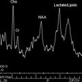
MR Imaging Workup of Inborn Errors of Metabolism of Early Postnatal Onset
Neonates with Seizures: What to Consider, How to Image
MR Imaging Workup of Inborn Errors of Metabolism of Early Postnatal Onset
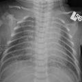 Birth-Related Injury to the Head and Cervical Spine in Neonates
MR Imaging of the Neonatal Musculoskeletal System
Birth-Related Injury to the Head and Cervical Spine in Neonates
MR Imaging of the Neonatal Musculoskeletal System
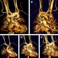 Congenital Cardiovascular Malformations: Noninvasive Imaging by MRI in Neonates
 MR Imaging of Neonatal Brain Infections
Stay updated, free articles. Join our Telegram channel

Full access? Get Clinical Tree


