and Jan Žižka2
(1)
Department of Radiology, University Hospital Hradec Králové, Hradec Králové, Czech Republic
(2)
Department of Radiology Faculty of Medicine in Hradec Králové, University Hospital Hradec Králové Charles University in Prague, Hradec Králové, Czech Republic
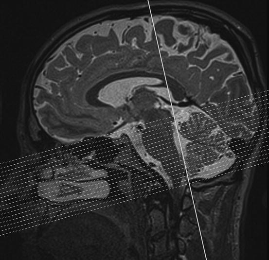
M00: MRI axial sections
Standard axial MRI section is perpendicular to the posterior margin of the brainstem defined on a midsagittal image.
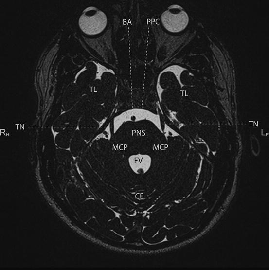


M01: MRI axial sections
BA
basilar artery
CE
cerebellum
FV
fourth ventricle
MCP
middle cerebellar peduncle
PNS
pons
PPC
prepontine cistern
TL
temporal lobe
TN
trigeminal nerve
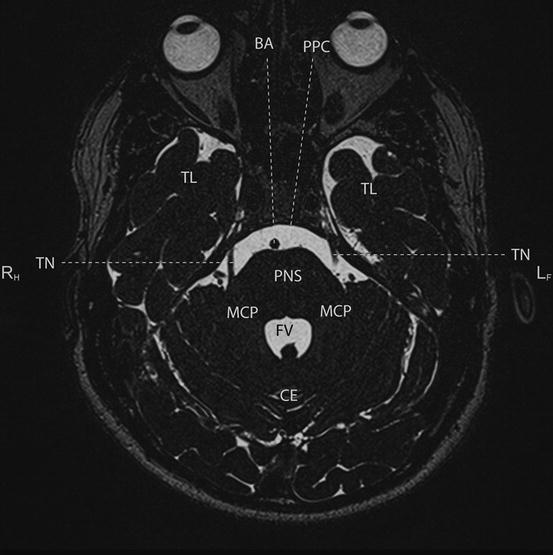
M02: MRI axial sections
BA
basilar artery
CE
cerebellum
FV
fourth ventricle
MCP
middle cerebellar peduncle
PNS
pons
PPC
prepontine cistern
TL
temporal lobe
TN
trigeminal nerve
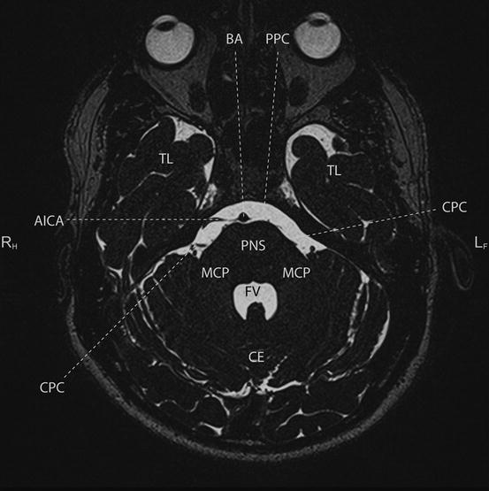
M03: MRI axial sections
AICA
anterior inferior cerebellar artery
BA
basilar artery
CE
cerebellum
CPC
cerebellopontine cistern
FV
fourth ventricle
MCP
middle cerebellar peduncle
PNS
pons
PPC
prepontine cistern
TL
temporal lobe
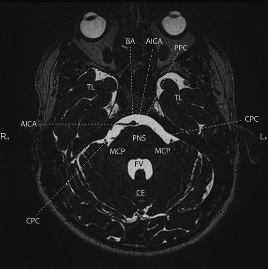
M04: MRI axial sections
AICA
anterior inferior cerebellar artery
BA
basilar artery
CE
cerebellum
CPC
cerebellopontine cistern
FV
fourth ventricle
MCP
middle cerebellar peduncle
PNS
pons
PPC
prepontine cistern
TL
temporal lobe
M05: MRI axial sections
AICA
anterior inferior cerebellar artery
BA
basilar artery
CE
cerebellum
CPC
cerebellopontine cistern
FV
fourth ventricle
MCP
middle cerebellar peduncle
PNS
pons
PPC
prepontine cistern
TL
temporal lobe
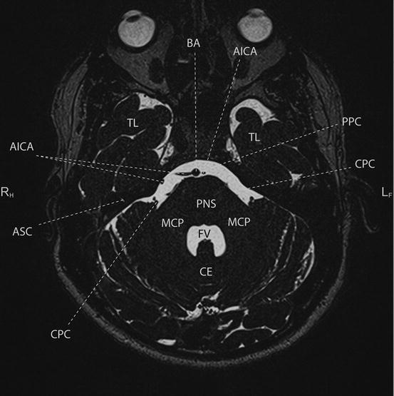
M06: MRI axial sections
AICA
anterior inferior cerebellar artery
ASC
anterior semicircular canal
BA
basilar artery
CE
cerebellum
CPC
cerebellopontine cistern
FV
fourth ventricle
MCP
middle cerebellar peduncle
PNS
pons
PPC
prepontine cistern
TL
temporal lobe
M07: MRI axial sections
AICA
anterior inferior cerebellar artery
ASC
anterior semicircular canal
BA
basilar artery
CE
cerebellum
CPC
cerebellopontine cistern
FV
fourth ventricle
MCP
middle cerebellar peduncle
PNS
pons
PPC
prepontine cistern
TL
temporal lobe
M08: MRI axial sections
AICA
anterior inferior cerebellar artery
ASC
anterior semicircular canal
BA
basilar artery
CE
cerebellum
CPC
cerebellopontine cistern
FV
fourth ventricle
MCP
middle cerebellar peduncle
PNS
pons
PPC
prepontine cistern
TL
temporal lobe
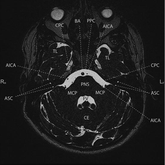
M09: MRI axial sections
AICA
anterior inferior cerebellar artery
ASC
anterior semicircular canal
BA
basilar artery
CE
cerebellum
CPC
cerebellopontine cistern
FV
fourth ventricle
MCP
middle cerebellar peduncle
PNS
pons
PPC
prepontine cistern
TL
temporal lobe
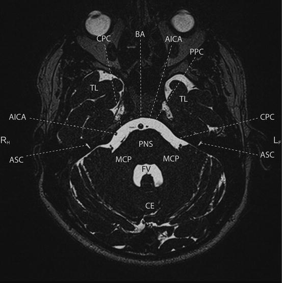
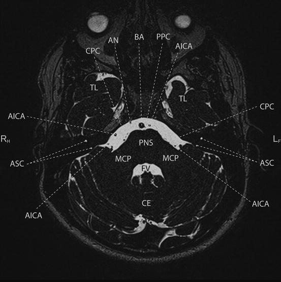
M10: MRI axial sections
AICA
anterior inferior cerebellar artery
AN
abducens nerve
ASC
anterior semicircular canal
BA
basilar artery
CE
cerebellum
CPC
cerebellopontine cistern
FV
fourth ventricle
MCP
middle cerebellar peduncle
PNS
pons
PPC
prepontine cistern
TL
temporal lobe
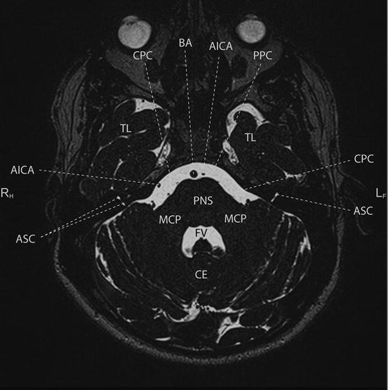
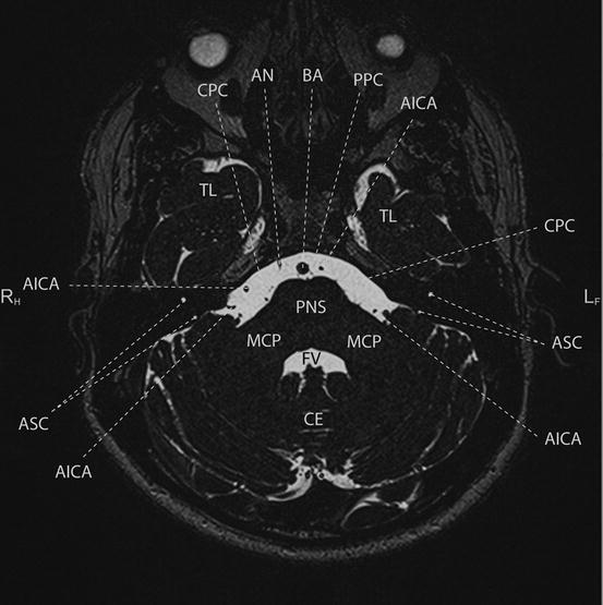
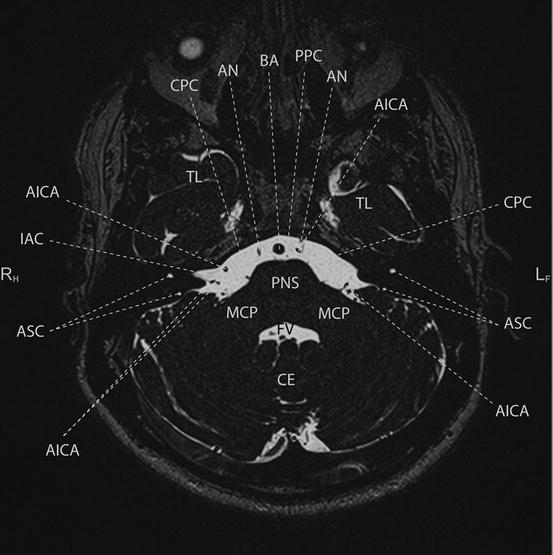
M11: MRI axial sections
AICA
anterior inferior cerebellar artery
AN
abducens nerve
ASC
anterior semicircular canal
BA
basilar artery
CE
cerebellum
CPC
cerebellopontine cistern
FV
fourth ventricle
MCP
middle cerebellar peduncle
PNS
pons
PPC
prepontine cistern
TL
temporal lobe
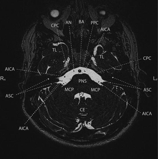
M12: MRI axial sections
AICA
anterior inferior cerebellar artery
AN
abducens nerve
ASC
anterior semicircular canal
BA
basilar artery
CE
cerebellum
CPC
cerebellopontine cistern
FV
fourth ventricle
MCP
middle cerebellar peduncle
PNS
pons
PPC
prepontine cistern
TL
temporal lobe
M13: MRI axial sections
Related posts:

Stay updated, free articles. Join our Telegram channel
Full access? Get Clinical Tree
