Table 9-1.
Muscles of the Hip
| MUSCLE | ORIGIN | INSERTION | NERVE SUPPLY |
|---|---|---|---|
| Obturator internus | Pelvic surface of the pubic rami near the obturator foramen, pelvic surface of the ischium between the foramen and the greater sciatic notch, deep surface of the obturator internus fascia, fibrous arch that surrounds the foramen for obturator vessels and nerve, most of the pelvic surface of the obturator membrane except the lower part | Medial side of the greater trochanter in front of the trochanteric fossa of the femur | Nerve to the obturator internus, from the lumbosacral trunk, and first and second sacral |
| Obturator externus | Lateral surface of the pubic and ischial rami, where they surround the obturator membrane, lateral surface of the obturator membrane | Trochanteric fossa | Obturator |
| Gemellus superior | Outer surface of the ischial spine and edge of the lesser sciatic notch | After union with the tendon of the obturator internus, inserts into the medial side of the greater trochanter in front of the trochanteric fossa | By a small nerve, branch of the nerve to obturator internus or branch of the nerve to the quadratus femoris |
| Gemellus inferior | Upper part of the inner border of the tuberosity of the ischium, sacrotuberous ligament, and edge of the lesser sciatic notch | By union with the tendon of the obturator internus or with the tendon onto the greater trochanter below the obturator internus muscle | By a small branch of the nerve to the quadratus femoris |
| Quadratus femoris | Upper part of the outer border of the tuberosity of the ischium | The inferior dorsal angle of the greater trochanter | Lumbosacral trunk and first sacral |
| Psoas major | By a series of thick fasciculi from the intervertebral discs and bodies between T12 and L5, from the bodies of L1 to L4, and from slender fascicles from the ventral surfaces of the transverse processes of the lumbar vertebrae | The lesser trochanter of the femur | Branches from L1 (often), L2, L3, and L4 |
| Iliacus | Iliac crest, iliolumbar ligament, iliac fossa, anterior sacroiliac ligaments, often from the ala of the sacrum, and from the ventral border of the ilium between the two anterior spines | Lateral surface of the psoas tendon (above the inguinal ligament) onto the femur immediately distal to the lesser trochanter; the lateral portion arises from the ventral border of the ilium and is attached to the tendon of the rectus femoris and the capsule of the hip joint | Femoral and L1 to L4 |
| Tensor fasciae latae | Anterior superior iliac spine and anterior part of the external lip of the iliac crest | Muscle fibers pass distally in parallel array, unite with the tendon, and join the iliotibial tract about one third of the way down the thigh | Superior gluteal |
| Gluteus medius | Ventral three fourths of the iliac crest, outer surface of the ilium between the anterior and posterior gluteal lines, and from the investing fascia | Onto the posterosuperior angle and the external surface of the greater trochanter | Superior gluteal (L4, L5, S1) |
| Piriformis | Lateral part of the ventral surface of S2, S3, and S4, posterior border of the greater sciatic notch, from the sacrotuberous ligament near the sacrum | Onto the anterior and inner parts of the upper border of the greater trochanter | S1 or S2 or from a loop between S1 and S2 |
| Gluteus maximus | Dorsal fifth of the outer lip of the iliac crest, ilium dorsal to the posterior gluteal line, thoracolumbar fascia between the posterior superior spine of the ilium and the side of the sacrum, lateral parts of S4, S5, and coccygeal vertebrae, and from the back of the sacrotuberous ligament | Into the iliotibial tract, gluteal tuberosity of the femur, adjacent part of the tendinous origin of the vastus lateralis | Inferior gluteal by two branches from the sacral plexus (separately or as a united nerve) |
| Gluteus minimus | Outer surface of the ilium between the anterior and inferior gluteal lines, from the septum between the gluteus minimus and the gluteus medius near the anterior superior, iliac spine and the capsule of the hip joint | Onto the anterior border of the greater trochanter of the femur | Superior gluteal from a branch that supplies the tensor fasciae latae |
| Biceps femoris, long head | Medial facet on the posterior surface of the ischial tuberosity and sacrotuberous ligament | By a tendon that extends to the head of the fibula | Tibial part of the sciatic |
| Semitendinosus | Distal margin of the ischial tuberosity and from the tendon common to it and the long head of the biceps femoris | By a triangular tendinous expansion into the proximal part of the medial surface of the tibia behind and distal to the insertion of the gracilis | Sciatic or directly from the lumbosacral plexus by two nerves: from S1 and S2 and from L5 and S1 |
| Semimembranosus | Lateral facet on the posterior surface of the ischial tuberosity | Posterior aspect of the medial tibial condyle | Sciatic branch (also supplies the adductor magnus) |
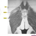
Axial
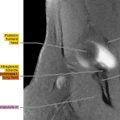
Figure 9.1.1

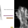
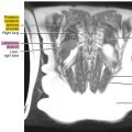
Figure 9.1.2

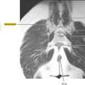
Figure 9.1.3

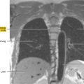
Figure 9.1.4

Figure 9.1.5
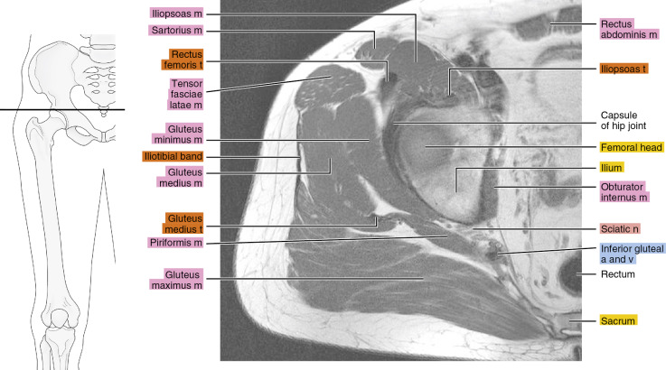
Figure 9.1.6

Figure 9.1.7

Figure 9.1.8

Figure 9.1.9

Figure 9.1.10

Figure 9.1.11

Figure 9.1.12