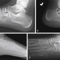
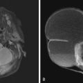
Whether the history is motor vehicle accident, fall from monkey bars, or sports-related injury, musculoskeletal trauma is one of the most common reasons for emergency department visits. Knowledge of the developing bone anatomy and specific pediatric fractures leads to appropriate diagnosis and treatment.
Common Musculoskeletal Trauma Imaging Techniques
Plain Radiographs
Multiorthogonal radiographs of the bones are a mainstay of trauma evaluation and are often the first line of evaluation.
Ultrasound
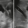
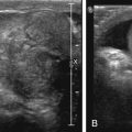
Ultrasound is often used in infants specifically for evaluation of cartilaginous portions of the bones. It is also used to assess for joint effusions, soft tissue lesions, or foreign body detection.
Computed Tomography
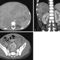
Complex fractures may necessitate additional evaluation by computed tomography (CT) to confirm/assess epiphyseal separation, comminution with fragments in the joint space, or the extent of fracture displacement or to determine for possible need of open reduction internal fixation. Contrast is occasionally used for evaluation of vascular injuries.
Magnetic Resonance Imaging
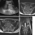
Magnetic resonance imaging (MRI) is an excellent modality for evaluation of soft tissue and osseous injuries or bone marrow edema or to detect avascular necrosis. Contrast is typically not necessary unless infection or neoplasm is considered.
Pediatric Bone Anatomy
The most important differences between evaluating pediatric and adult bones are the anatomy of the developing bone, including the presence of growth plates, apophyses, and ossification centers.
Approximately 15% of extremity fractures in children involve disruptions of the growth plate, which is two to five times weaker than any other structure in the pediatric skeleton. In addition, the pediatric bone has greater porosity than mature adult bone, explaining unique pediatric fractures: plastic deformation, buckle, and greenstick fractures. Its greater porosity is secondary to its increased vascularity, which decreased with increasing age.
Long bones anatomy in pediatric patients include ( Fig. 17.1 ):
Epiphysis: This is located at the end of the bone between the physis and the joint space.
Physis (growth plate): The primary physis is made of hyaline cartilage and is responsible for the longitudinal growth via endochondral ossification. The newest bone forms on the metaphyseal side of the physis.
Metaphysis: This is a flared, highly vascularized segment of bone between the diaphysis and physis. It is made of immature bone. Its thinner cortex relative to the diaphysis is in part owed to its greater vascularity.
Metadiaphysis: A segment of bone between the diaphysis and metaphysis, where there is a transition in cortical thickness from the diaphyseal thicker cortex and the thinner metaphyseal cortex, increasing its susceptibility to fracture.
Diaphysis: The shaft between the proximal and distal metaphysis. The diaphysis has a thicker cortex than the metaphysis.
Apophysis: Bone that arises from a separate ossification than the parent bone and eventually fuses with the parent bone. It does not contribute to longitudinal growth and often has a tendinous attachment ( Table 17.1 ).
TABLE 17.1
Pelvis apophyses and their tendinous attachments
Apophysis
Tendon
Anterior superior iliac spine
→
Sartorius
Anterior inferior iliac spine
→
Rectus femoris
Lesser trochanter
→
Iliopsoas
Greater trochanter
→
Gluteus medius
Ischial apophysis
→
Hamstring
Iliac crest
→
Abdominal wall muscles

Particularly Pediatric Fractures
Plastic Deformation
This fracture is caused by compressive trauma severe enough to deform a bone without a radiographically evident fracture. Plastic deformation is most often seen in the radius, ulna, and fibula. Although a fracture line is not visible, subsequent radiographs may show periosteal reaction as a result of fracture healing. Due to disruption of the “ring” in the forearm or distal lower extremity, an additional fracture may be present in the companion bone ( Fig. 17.2 ).

Buckle
A buckle fracture is an incomplete fracture at the junction of the metaphysis and diaphysis on the compressive side of the bone resulting in a focal bulge on radiographs ( Fig. 17.3 ). There is no discrete fracture line, and the bulge does not extend along the circumference of the bone. These fractures are commonly seen in the distal radius and ulna, secondary to a fall on an outstretched hand.

Greenstick Fracture
A greenstick fracture is an incomplete cortical disruption along the tension side of bone, with a plastic deformation on the compressive side, usually occurring at the metadiaphysis ( Fig. 17.4 ).

Complete Fracture
A complete fracture is one that propagates through the bone resulting in a circumferential disruption of the cortex.
Physeal Fractures
The Salter-Harris classification is the most commonly used classification system to describe fractures involving the physis ( Fig. 17.5 ) ( Table 17.2 ). Approximately 90% of growth plate fractures can be classified as Salter-Harris I–IV on plain radiograph. The other 10% of fractures may require either additional views or advanced imaging for diagnosis. Although the majority of growth plate injuries heal without complication, limb length discrepancies or angular deformities are often the result of trauma to the physis, causing injury to the physis itself or disruption of the epiphyseal or metaphyseal blood supply and subsequent growth arrest and physeal bar development.


Stay updated, free articles. Join our Telegram channel

Full access? Get Clinical Tree


