Chapter 3
Multiple Clusters of Crushed Stone-like Calcifications on the Mammogram Produced by Malignant Processes
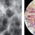
Mammograms showing multiple clusters (or rarely a single cluster) of crushed stone-like calcifications that represent only a portion of the underlying disease can be subdivided by careful correlation of mammographic; large-section histological; and subgross, thick-section histological images into the following two subgroups. Group 2A The in situ process is primarily confined to TDLUs that are separated from each other by normal tissue, although histology may reveal a few involved ducts. In these cases there is reasonable concordance between the mammographic and histological findings. The crushed stone-like calcifications may also be associated with powdery calcifications, seen in close proximity on the mammograms, representing Grade 2 and Grade 1 in situ carcinoma side by side. Group 2B Although the mammographic image may show multiple clusters which appear similar to those in Group 2A, large-section histological examination demonstrates tightly packed cancerous TDLUs and ducts covering a larger area than that indicated by the calcifications detected at mammography. MRI of the breast plays an important role in revealing the true extent and confluent nature of the underlying disease. In some extreme cases very large numbers of clusters of crushed stone-like calcifications are visible on the mammogram, often associated with some casting type calcifications. The larger the number of clusters, the more difficult it is to discern the individual clusters. Correlation with large-section and subgross, 3D histology shows a high density of tightly packed cancerous TDLUs and ducts with little, if any, normal tissue in between. Histological signs of neoductgenesis and lymph vessel invasion are often present and may account for the occasional poor outcome.
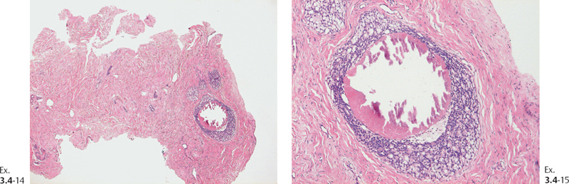
Group 2A: Multiple Foci on Histology with Intervening Normal Tissue
Example 3.1
A 62-year-old asymptomatic woman, screening examination.
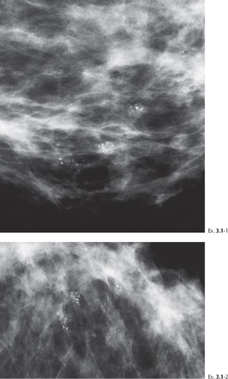
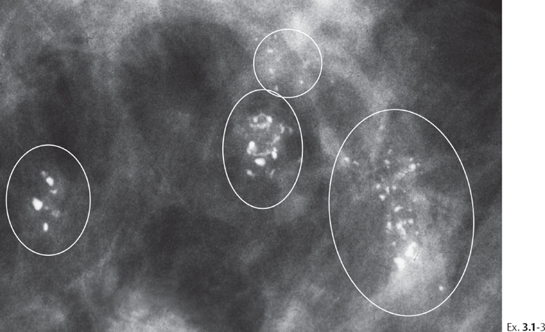
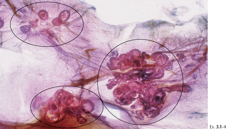
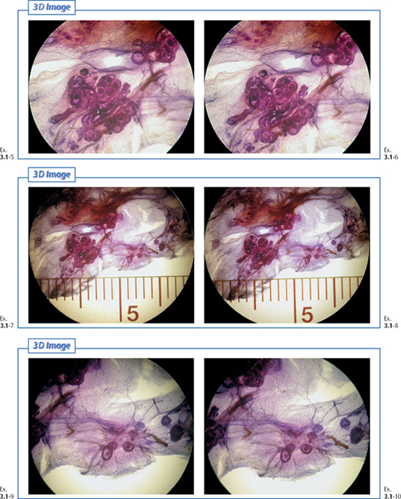
Treatment and outcome: Sector resection and postoperative irradiation. The patient died of acute leukemia 12 years after treatment. During the follow-up period there was no evidence of breast cancer recurrence.
Example 3.2
A 55-year-old asymptomatic woman, screening examination. She was called back for further assessment of the calcifications detected on the screening mammograms.
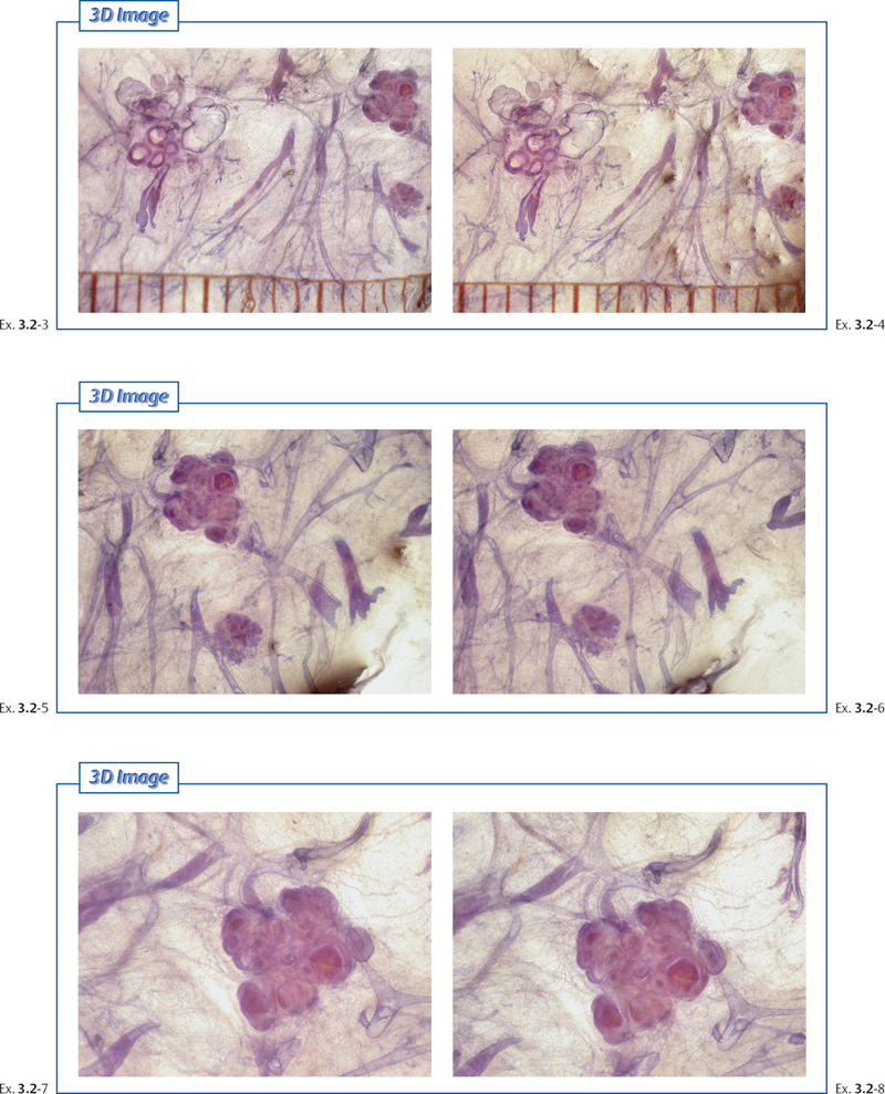
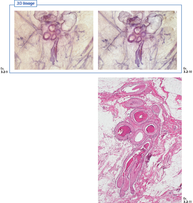
Final histology: Grade 2 in situ carcinoma over an area measuring 50 mm in diameter. Cancer cells were demonstrated at the resection margin.
Treatment and outcome: Sector resection. Histological examination has found cancer foci on the margin. Mastectomy: no residual tumor, pNX. The patient was symptom-free at the most recent follow-up examination 12 years after operation.
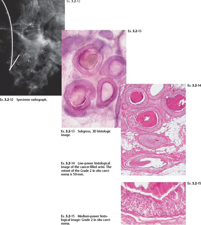
Comment
The multiple clusters on the mammogram reflect the multiple cancerous TDLUs. Unlike the contiguous tumor growth characteristic of casting type calcification cases and the subgroup 2B, the multiple cluster crushed stone-like calcification cases described at the beginning of this chapter originate in individual TDLUs that are separated from each other by a few millimeters of intervening normal tissue. Viewing the large thick-section/3D histological slices not only provides excellent correlation with the mammographic findings but also unearths additional, valuable information, such as mammographically occult cancer foci. In this particular example, the distance between two of the calcified TDLUs was 8 mm. These observations suggest that the term “close” surgical margin may be an unreliable description when the finding on the mammogram consists of multiple clusters of crushed stone-like calcifications.
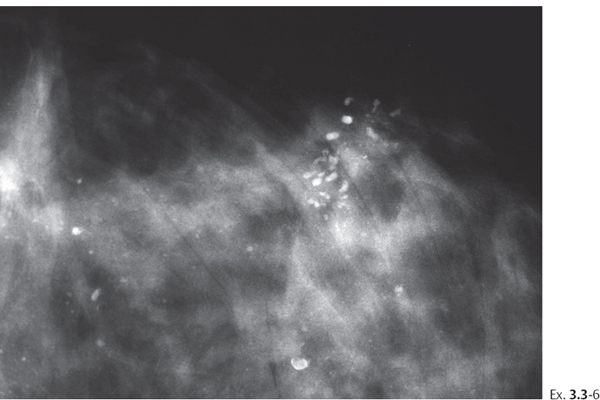
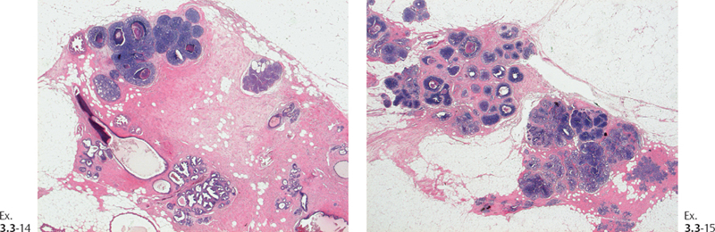
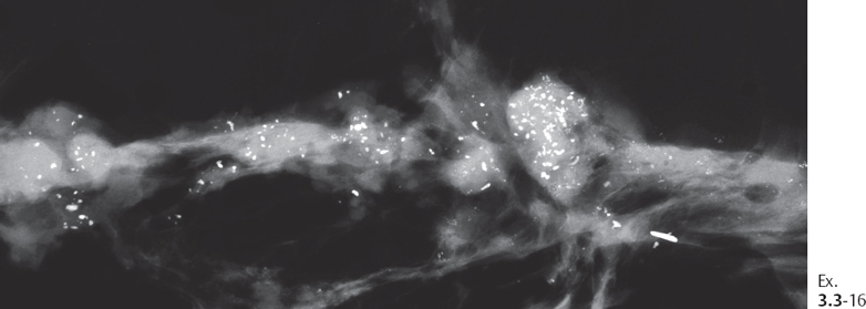
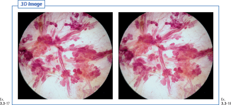
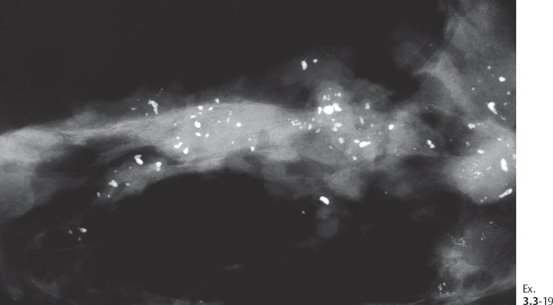
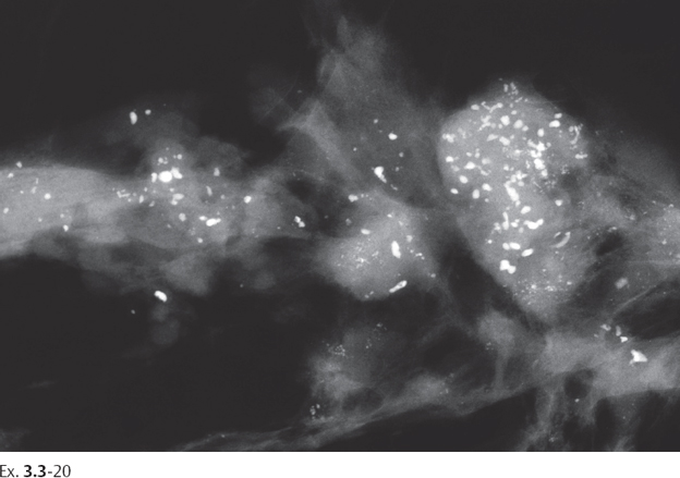
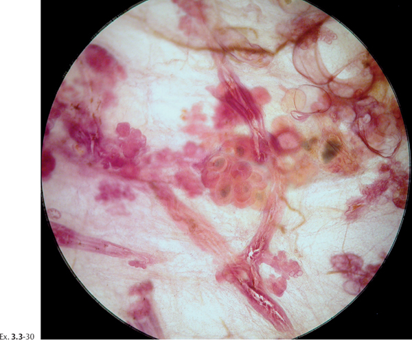
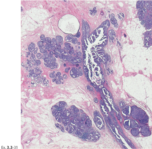
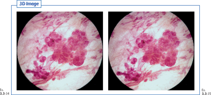
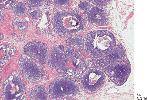
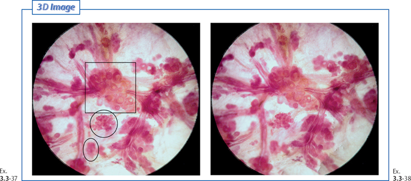
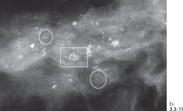
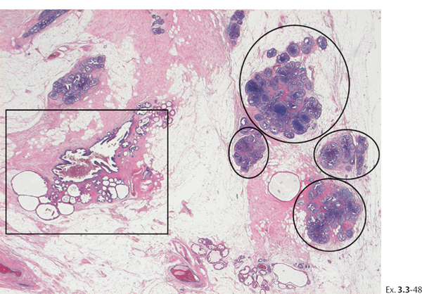
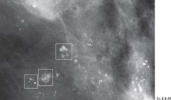
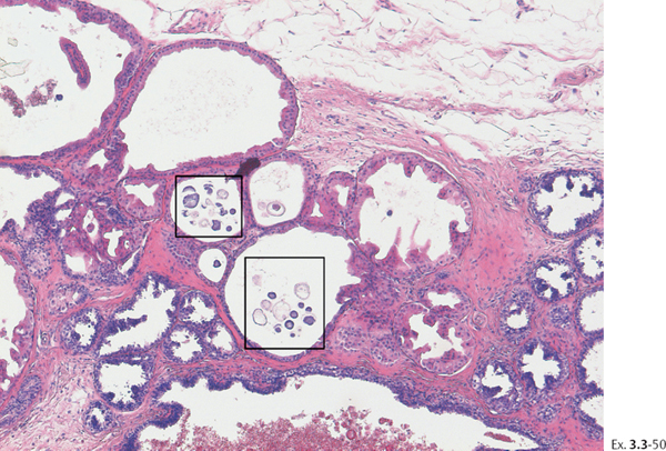
Example 3.3
This 65-year-old woman was called back from mammography screening for assessment of multiple clusters of calcifications in the right breast.
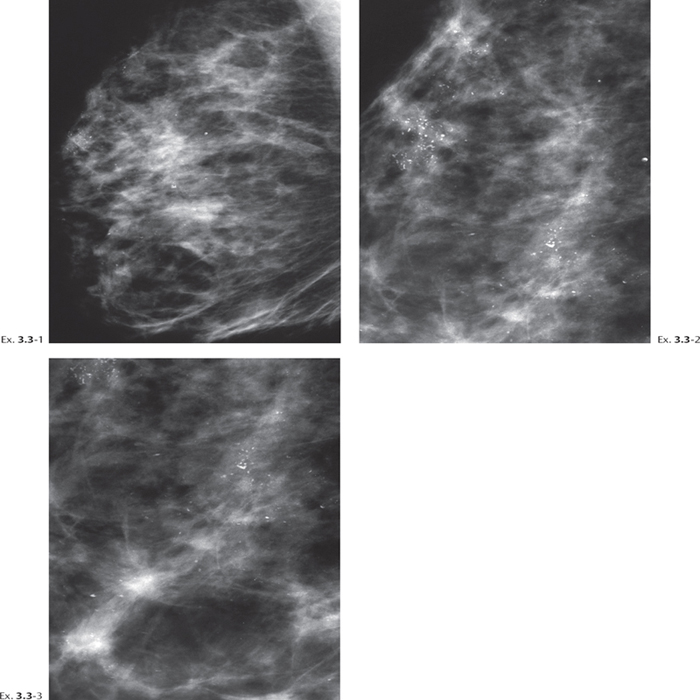
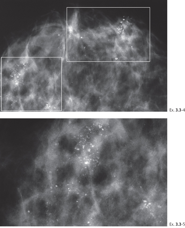
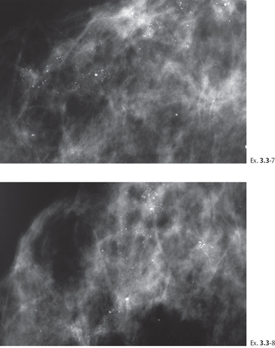
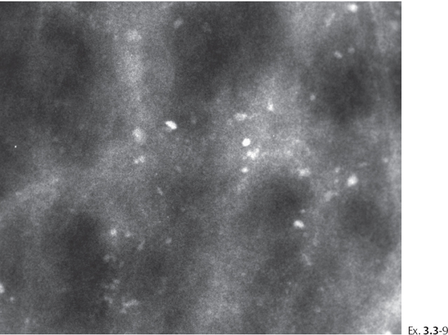
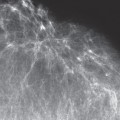
Ex. 3.3-7 to 9 Right breast, CC projection, microfocus magnification images at increasing enlargement.
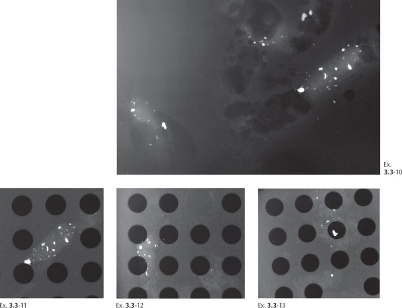
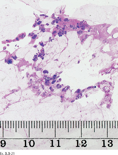
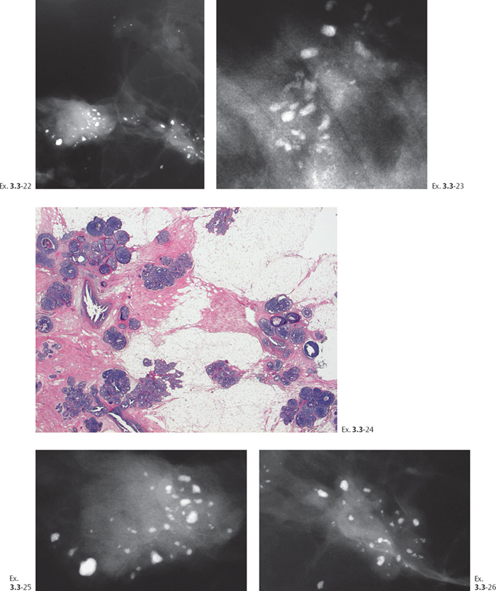
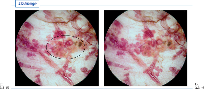
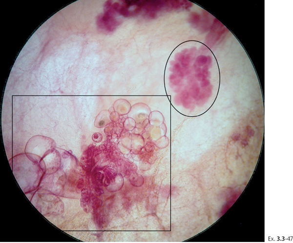
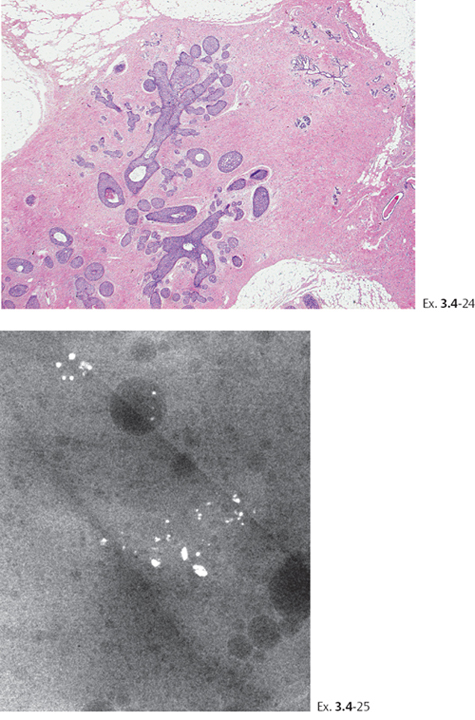
Ex. 3.3-22 to 26 Histological-mammographic correlation of the multiple clusters of cancerous TDLUs containing the crushed ston-elike calcifications

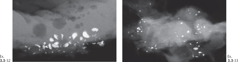
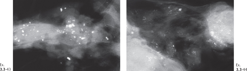
Ex. 3.3-32 & 33 Specimen radiographs of multiple clusters of crushed stone-like, pleomorphic calcifications.
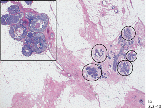
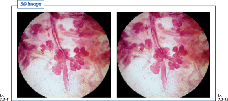
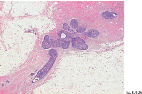
Ex. 3.3-41 & 42 Subgross histological image pair of several TDLUs and a subsegmental duct distended by in situ carcinoma.
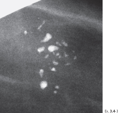
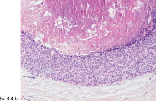
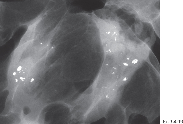
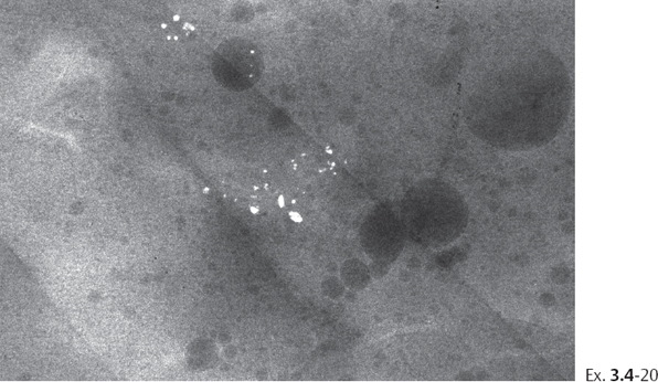
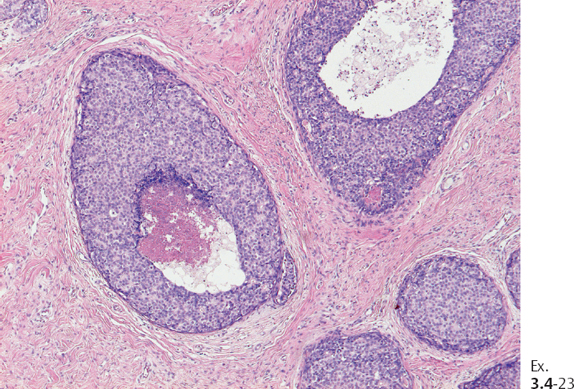
Example 3.4
A 54-year-old asymptomatic woman, screening examination. Her sister had breast cancer at age 59 years.
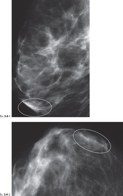
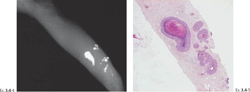
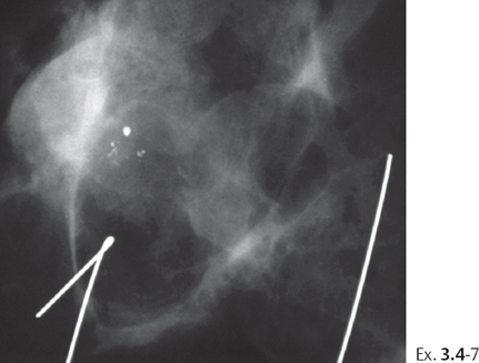
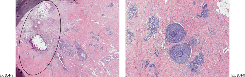
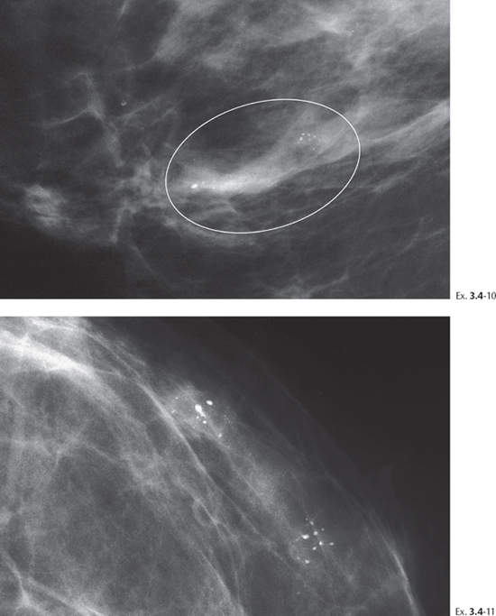
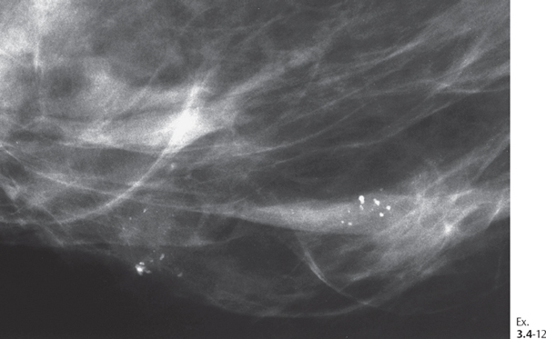
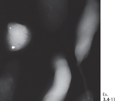
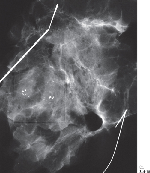
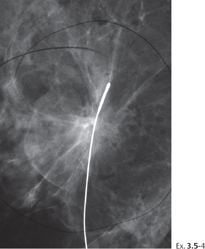
Ex. 3.4-16 Operative specimen radiograph following preoperative localization using the bracketing technique.
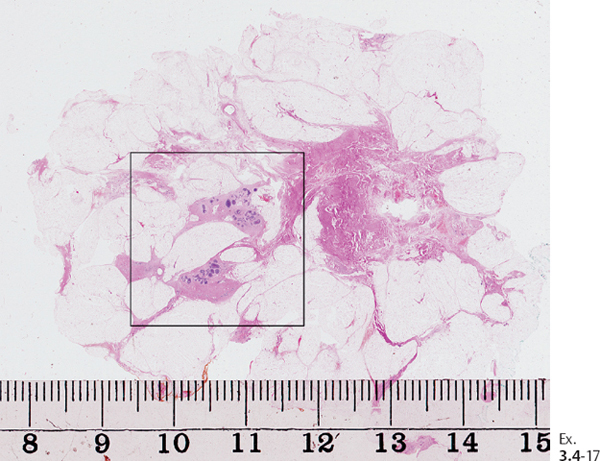
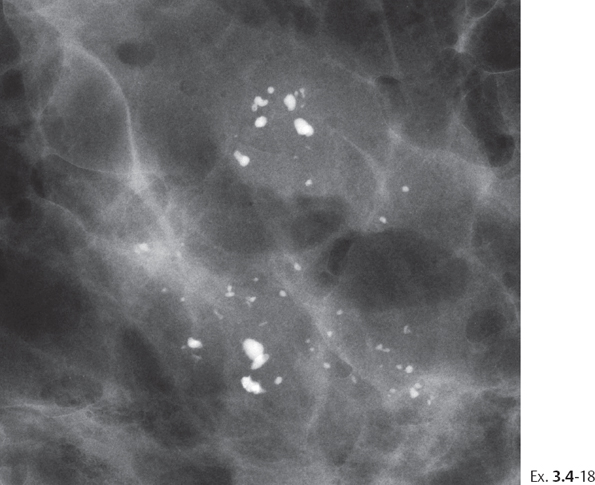
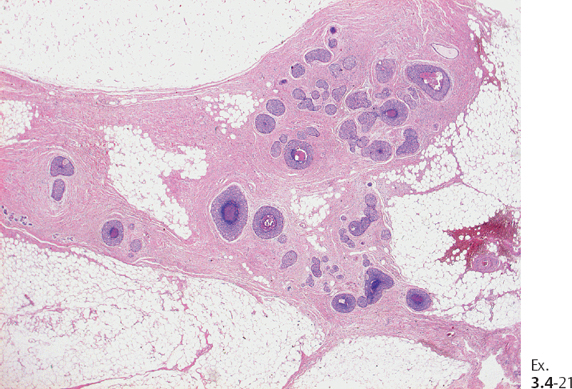
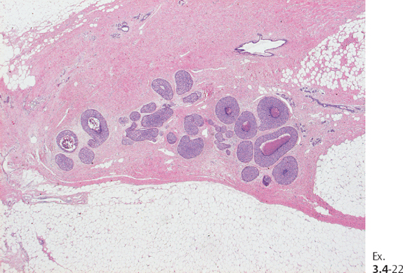
Treatment and outcome: Sector resection and postoperative irradiation following the diagnosis of recurrence. The patient had no evidence of disease at the most recent follow-up examination 2 years after her second operation.
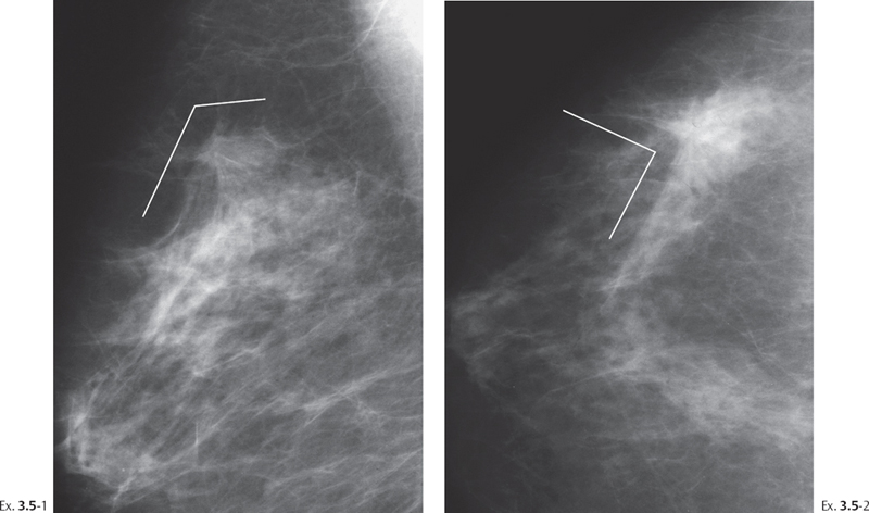
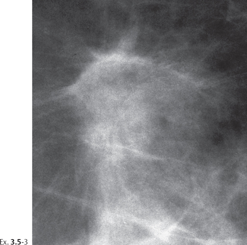
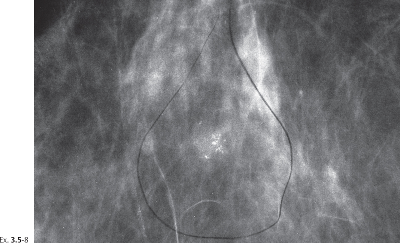
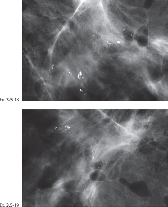
Example 3.5
A 57-year-old asymptomatic woman, screening examination.
Follow-up examination 6 years after surgery.
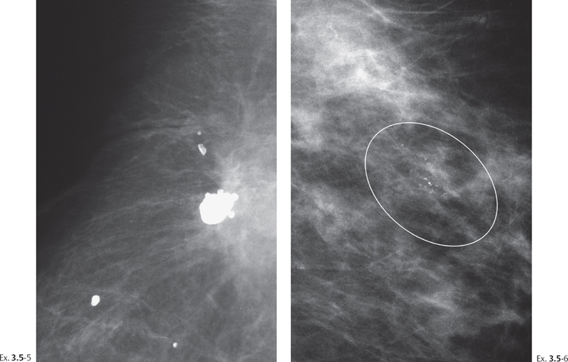
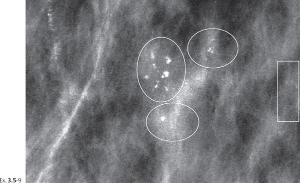
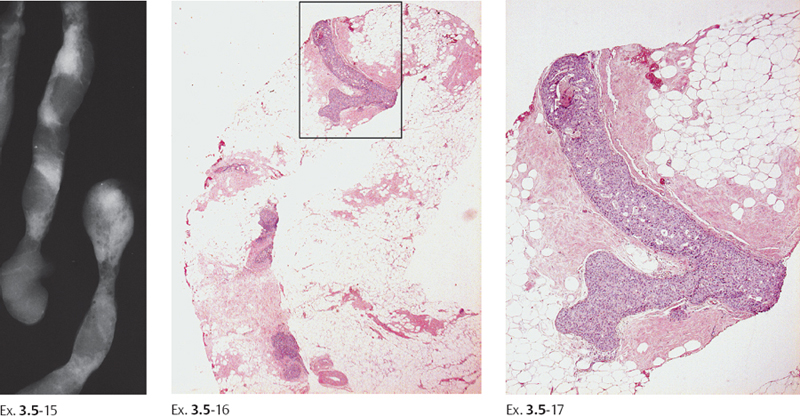
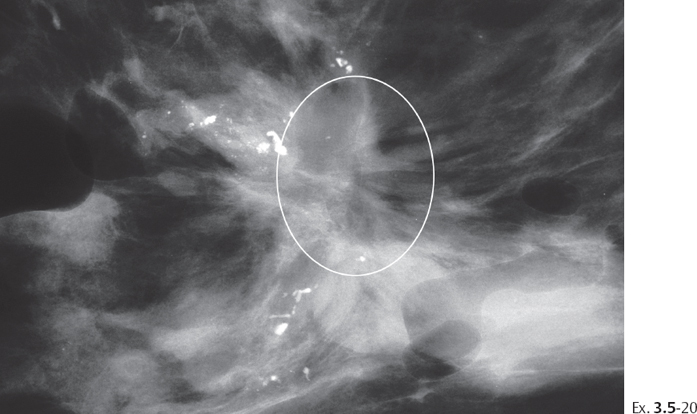
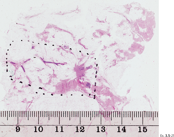
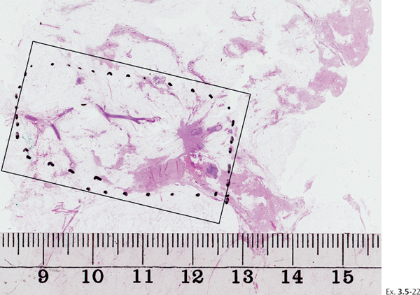
Ex. 3.5-22 The large-section histology image can be compared with the detailed photomicrographs on these two pages (Ex. 3.5-23 to 29).
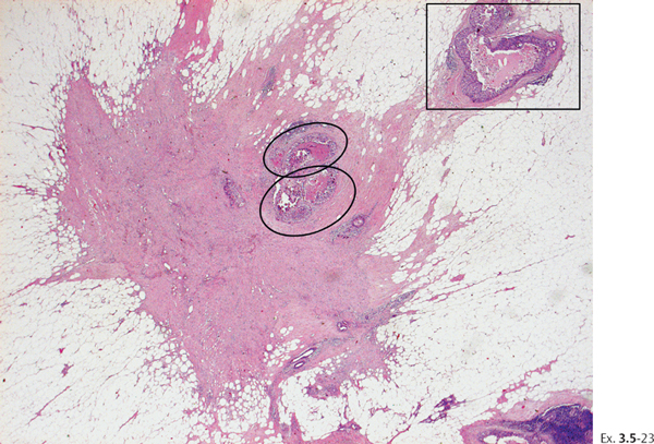
Ex. 3.5-23 The star-shaped lesion (encircled on Ex. 3.5-22) is a scar caused by the preoperative needle biopsy performed 3 weeks earlier. There are in situ carcinoma foci (ovals) entrapped within the scar. Another in situ focus is outlined by a rectangle.
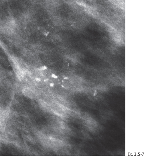
The photomicrographs on this page are higher-power views of the in situ foci within the rectangle on Ex. 3.5-22.
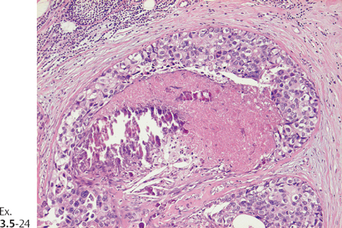
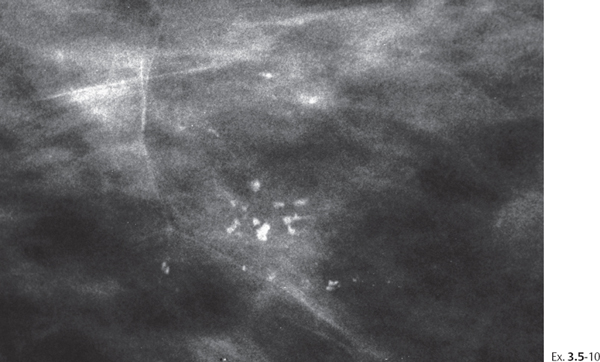
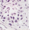
Ex. 3.5-24 High-power magnification of one of the in situ foci (upper oval on Ex. 3.5-23) with solid cell proliferation, central necrosis, and amorphous calcification.
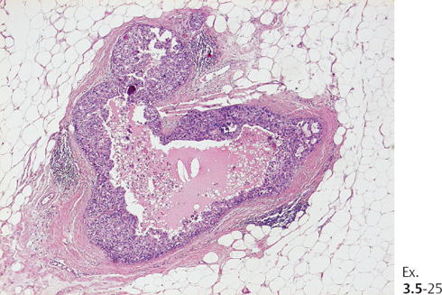
Ex. 3.5-25 Higher magnification of another focus of in situ carcinoma, seen on Ex. 3.5-23 within the rectangle.
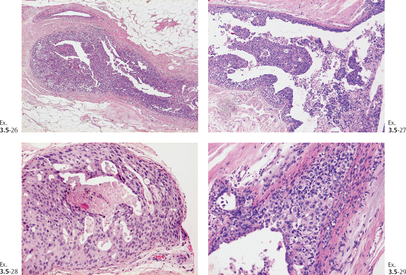
Ex. 3.5-26 to 29 Four enlarged images of the noncalcified in situ carcinoma foci within the rectangle on Ex. 3.5-22. The area with in situ carcinoma measured 40 mm ⊠ 15 mm, considerably larger than the area with crushed stone-like calcifications on the mammogram.
Treatment and outcome: Right breast: sector resection and postoperative irradiation. Left breast (6 years later): mastectomy. The patient had no signs of recurrence 2 years following mastectomy of the left breast.
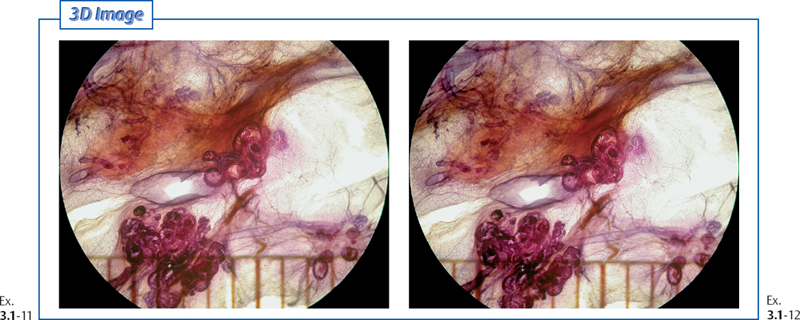
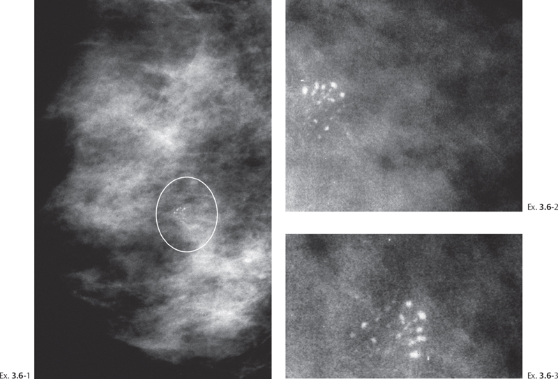
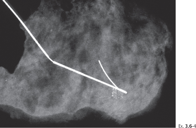
Example 3.6
A 47-year-old asymptomatic woman, screening examination. She was called back for further assessment of the small cluster of calcifications detected on the screening mammograms.
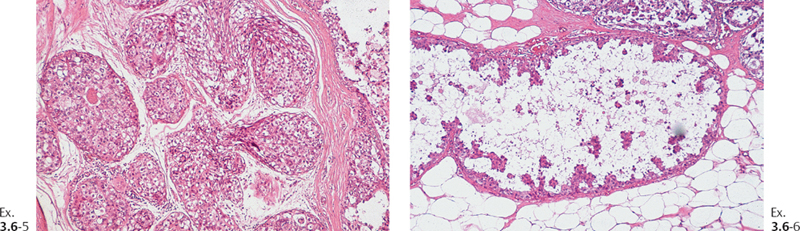
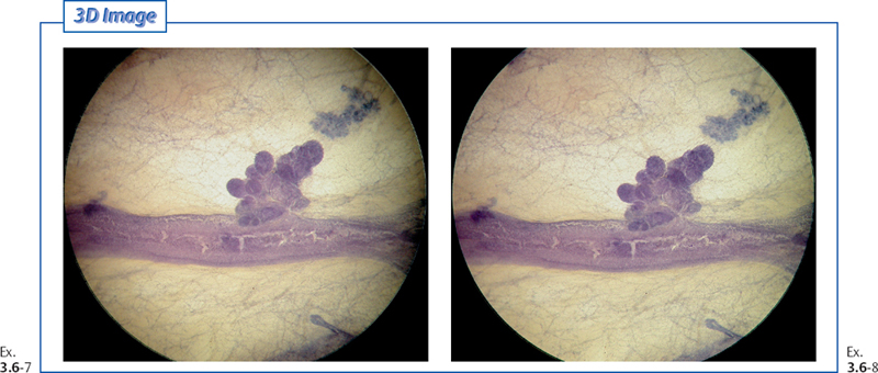
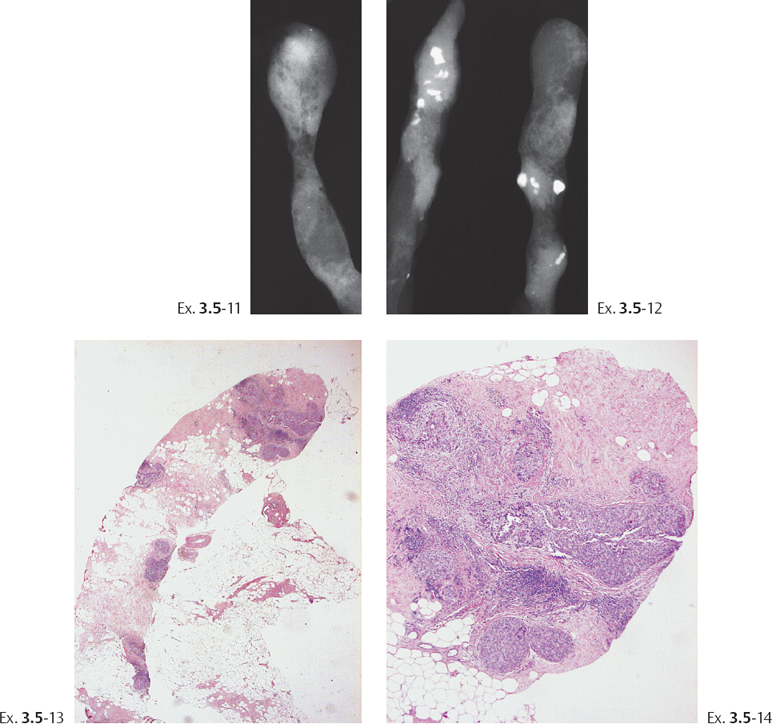
Ex. 3.6-7 & 8 Stereoscopic subgross 3D images of a section of a cancerous duct and an associated TDLU with in situ carcinoma. The conventional histological image of this TDLU is seen in Ex. 3.6-5.
Ex. 3.6
Related posts:
 The Prognostic Importance of Mammographic Tumor Features
 The Diagnostic Approach to Malignant Type Calcifications on the Mammogram
 Differential Diagnosis of Breast Diseases Producing Clustered, Discernible Calcifications
 Group 1: One or Two Clusters of Crushed Stone-like Calcifications of the Mammogram Produced by Malignant Processes
 Local Staging: Imaging Options and Core Biopsy Strategies
 Calcifications: Eggshell/Rim
Stay updated, free articles. Join our Telegram channel

Full access? Get Clinical Tree


