Muscle Injury
Get Clinical Tree app for offline access
IMAGING
General Features
Ultrasonographic Findings
 Typically associated with grade 2 or 3 tears
 Appearance varies with duration, severity, and location of hematoma
 During first 48 hours, initially hypoechoic then becomes more hyperechoic as hematoma solidifies
 After 72 hours, hematoma can start to liquefy
 Marginal hyperemia during reparative phase (reparative fibrovascular granulation tissue)
Imaging Recommendations


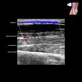
 at the distal end of the medial belly of gastrocnemius muscle. This is the most common site of calf muscle tears.
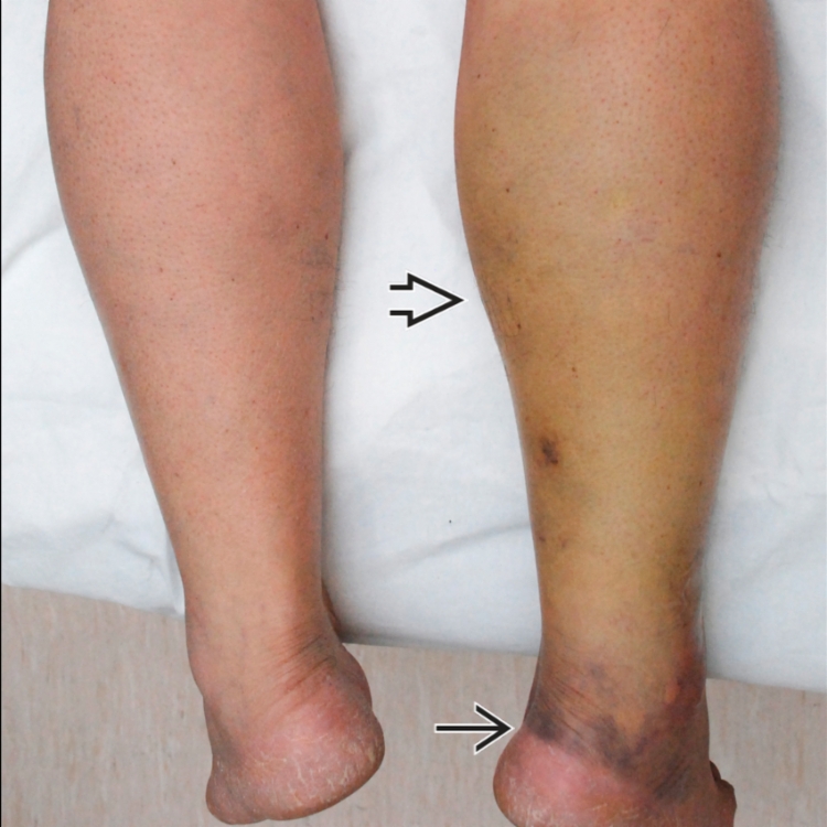
 with more intense, gravity-related bruising at the ankle region
  .
.
 at the distal end of the medial belly of gastrocnemius muscle. The medium-severity tear is filled with fluid. No muscle retraction is present.
 . Overall, ~ 15% of the cross-sectional area (CSA) of the muscle insertion is torn. The plantaris tendon
  is normal.