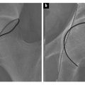
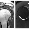
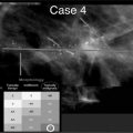
Fig. 1
Hip joint of a 6-year-old boy. Normal findings. Arrows point to growth plates. apo apophysis, epi epiphysis, meta metaphyis
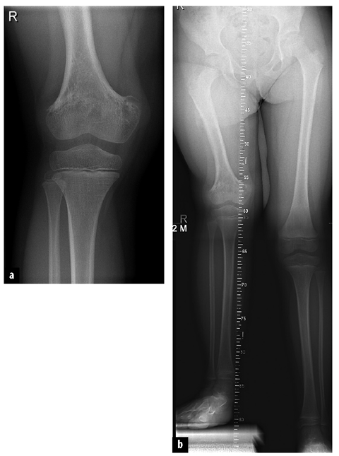
Fig. 2 a, b.
7-year-old girl developed premature closure of the growth plate of the distal femur (a) after ostemyelitis at the age of 8 months, resulting in severe leg-length discrepancy (b)
On the other hand, overgrowth (both enchondral and membranous growth) occurs in diseases that increase blood flow to the growth plate during a prolonged period of time. This phenomenon is present in vascular malformations and juvenile idiopathic arthritis. Adults with similar diseases (rheumatoid arthritis) present with radiological features other than overgrowth.
Growth lines (synonyms: growth recovery lines, growth arrest lines, Park-Harris lines) are thin sclerotic metaphyseal lines parallel to the growth plate. Usually they represent short periods of growth arrest during which the newly formed osteoid remains longer than usual in the zone of provisional calcification. These lines are nonspecific and can be caused by trauma, systemic illness and chemotherapy. Often no cause can be found (idiopathic). Growth lines can also be formed during short periods of hypermineralization, such as in vitamin D overload or bisphosphonate therapy (in patients with osteogenesis imperfecta). Lead bands in chronic lead intoxi cation are, in a way, caricatures of growth lines.
Growth lines disappear during growth, and can persist into adulthood, but they will not appear during adulthood.
During childhood, growth plates progressively become smaller until they close and the epiphysis and metaphysis fuse. However, some diseases simulate a widening of the growth plate (e.g., rickets, see section below on metaphysis).
Metaphysis
Metaphyses are unique for the pediatric skeleton; after closure of the growth plate the metaphysis and epiphysis fuse and cease to exist as separate structures.
The metaphysis is characterized by high metabolism and vascularization; this is to be expected because both mineralization and enchondral bone formation take place at this location. In a sense, the metaphysis is the mirror of bone metabolism. All systemic diseases that alter bone metabolism will radiologically first become evident at the metaphysis. For instance, rickets (dietary, renal or hereditary) affects the whole skeleton but is radiologically recognized by its typical metaphyseal changes (fraying, splaying and cupping). Lead intoxication shows characteristic metaphyseal bands of increased density, whereas childhood leukemia demonstrates lucent metaphyseal bands (Fig. 3). In adults, these diseases show much less conspicuous skeletal changes and are more difficult to detect radiologically.
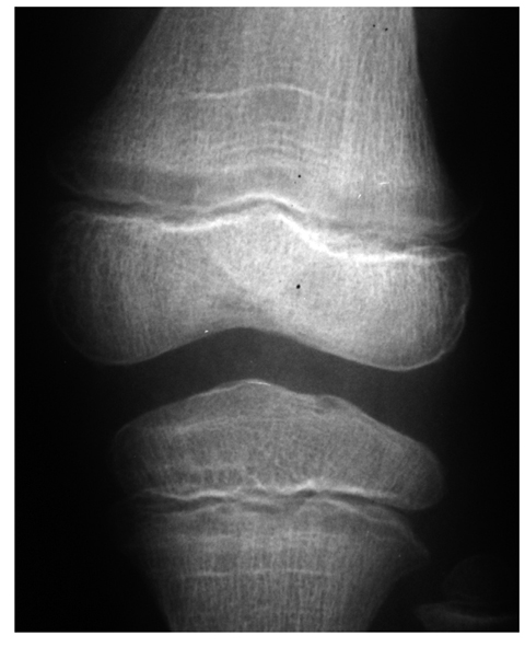

Fig. 3
A 5-year-old boy with a limp. Lucent metaphyseal bands at the proximal tibia and distal femur. Similar abnormalities were found at the hip. Diagnosis was acute lymphatic leukemia
Because the metaphyseal vessels terminate in slow flow venous sinusoidal lakes, the metaphysis is predisposed as the starting point for acute hematogenous osteomyelitis [4]. The manifestation of osteomyelitis in children is age dependent. In infants, diaphysial vessels penetrate the growth plate to reach the epiphysis, facilitating epiphysial and joint infections in this age group [5, 6].
In older children, the growth plate constitutes a barrier for the diaphyseal vessels; in adults, the epiphysis and metaphysis each have a separate blood supply and infection from bone to joint is far less common [7].
Epiphysis
Epiphyses are unique for the pediatric skeleton; after closure of the growth plate the epiphysis and metaphysis fuse and cease to exist as separate structures.
In contrast to the metaphysis, the epiphysis has a low metabolism and therefore little vascularization. So, from a metabolic point of view, the epiphysis is an uninteresting structure. However, there are some features that make the epiphysis radiologically interesting.
First, all epiphyses form joints, and the carpal and tarsal bones can also be considered as epiphyseal structures forming joints. Therefore diseases of epiphyseal structures can lead to severely disabling joint abnormalities. Structures that are anatomically and histologically similar but do not form joints are called apophyses (e.g., the greater trochanter; Fig. 1).
Second, these structures have a poor vascularization, not only because of their low metabolism but also because the growth plate forms an absolute barrier for blood vessels. This makes epiphyses, and carpal and tarsal bones prone to avascular necrosis. Many of these conditions have specific names, as shown in Table 1.
Table 1


Avascular necrosis
Stay updated, free articles. Join our Telegram channel

Full access? Get Clinical Tree


