, Maria Custódia Machado Ribeiro2 and Bruno Beber Machado3
(1)
Hospital da Criança de Brasília – Jose Alencar, Clínica Vila Rica, Brazília, Brazil
(2)
Hospital da Criança de Brasília – Jose Alencar, Hospital de Base do Distrito Federal, Brasília, Brazil
(3)
Clínica Radiológica Med Imagem, Unimed Sul Capixaba, Santa Casa de Misericordia de, Cachoeiro de Itapemirim, Brazil
Abstract
The vast majority of hematologic diseases (hemoglobinopathies, coagulopathies, or malignancies) lead to some kind of osteoarticular involvement. Such involvement is often responsible either for the clinical presentation or for some of the most disabling features of these conditions. It is not uncommon for the radiologist to be the one who first suggests the diagnosis. Awareness of the imaging appearance of the main pediatric hematologic diseases is essential in order to avoid diagnostic errors and allow timely institution of treatment.
8.1 Introduction
The vast majority of hematologic diseases (hemoglobinopathies, coagulopathies, or malignancies) lead to some kind of osteoarticular involvement. Such involvement is often responsible either for the clinical presentation or for some of the most disabling features of these conditions. It is not uncommon for the radiologist to be the one who first suggests the diagnosis. Awareness of the imaging appearance of the main pediatric hematologic diseases is essential in order to avoid diagnostic errors and allow timely institution of treatment.
8.2 Hemoglobinopathies
The hemoglobinopathies encompass a heterogeneous group of diseases in which there is some abnormality of the hemoglobin molecule, frequently associated with hemolytic anemia. The most prevalent of them is sickle-cell disease (SCD), an autosomal recessive condition that predominates in Afro-descendants. In SCD, there is an abnormal form of hemoglobin with a mutant beta globin chain, named hemoglobin S; it predominates over hemoglobin A, which is the dominant form in healthy individuals. Upon deoxygenation, erythrocytes with hemoglobin S lose their malleability and undergo irreversible deformation (sickling), being removed from circulation by the reticuloendothelial system. Thalassemia is the most frequent hemoglobinopathy in individuals with Mediterranean, Arab, or Asian ancestry, characterized by defective formation of alpha or beta chains of the hemoglobin molecule, being the latter (beta-thalassemia) the most common form. Because of its prevalence, SCD will be used as the prototype to describe the imaging findings of hemoglobinopathies unless otherwise specified.
Chronic anemia related to SCD leads to persistence and hyperplasia of the hematopoietic red marrow (see Chap. 2 – Fig. 8.1), with osteoporosis, widening of the medullary cavities, coarse appearance of bone trabeculae, and thinning of the cortex (Fig. 8.2). Classic radiographic findings in the calvaria include widening of the diploic space, thickening of the inner table, thinning of the outer table, and prominent trabeculae (hair-on-end sign) (Fig. 8.3). Spinal involvement is characterized by biconcave “fish” (or “fish mouth”) vertebrae, cortical thinning, and weakening of the medullary bone, all related to osteoporosis, with vertebral wedging/collapse in advanced cases (Figs. 8.4 and 8.5).
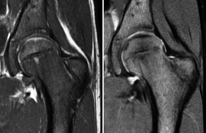
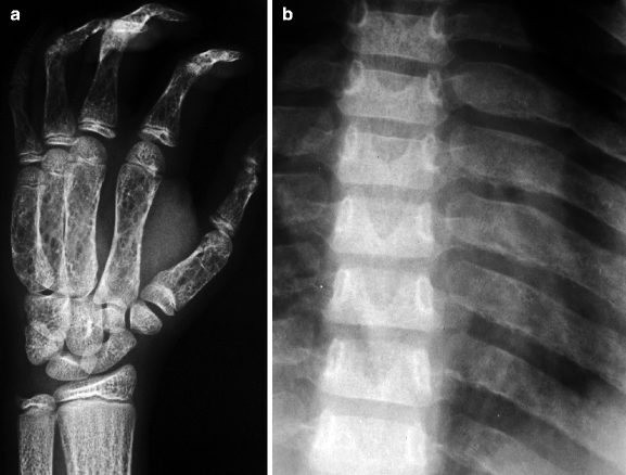
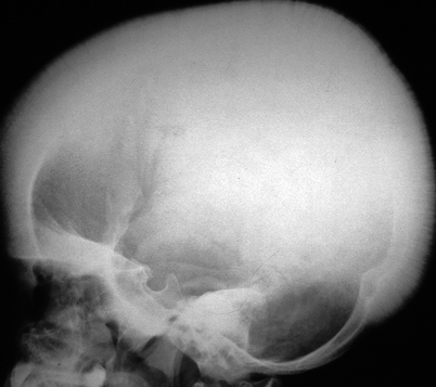
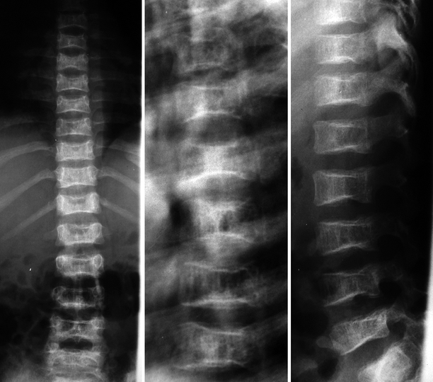
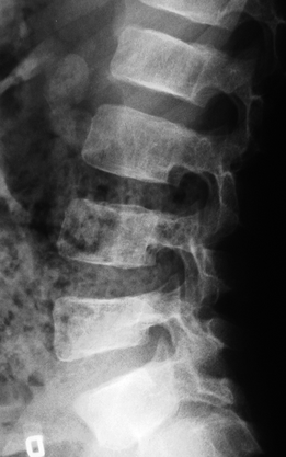
Fig. 8.1
Coronal T1-WI (left) and fat sat T2-WI (right) of the left hip of a child with SCD. There is diffuse alteration of the signal intensity of the bone marrow, which presents low/intermediate signal intensity on T1-WI and hyperintense on fat sat T2-WI, representing hyperplasia of the red marrow related to chronic anemia
Fig. 8.2
Radiographic abnormalities related to marrow hyperplasia in SCD. In (a), there is enlargement of the tubular bones of the right hand and coarse appearance of the trabeculae. In (b), the ribs are widened due to enlargement of the medullary cavities, with irregular periosteal reaction in several of them at left
Fig. 8.3
Lateral radiograph of the skull of a 7-year-old child with SCD. Widening of the diploic space is evident, with prominence of bone trabeculae, thickening of the inner table, and thinning of the outer table. The occipital squama is spared
Fig. 8.4
Radiographic abnormalities of the spine of a child with SCD. There is diffuse osteoporosis and thickening of the trabeculae. Several vertebrae present mildly reduced height and biconcave appearance, notably in the dorsal spine, related to hyperplasia of red marrow. “H-shaped” vertebrae are also present in the lumbar spine
Fig. 8.5
Lateral view of the lumbar spine of a child with SCD revealing diffuse osteoporosis and reduction of the height of L2 and L5, as well as subtle posterior wedging of the latter
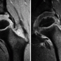
Abnormalities of bone development are also common in children with SCD, mostly related to ischemia of the growth cartilages (vasoocclusion due to trapping of the abnormally shaped erythrocytes). In the ankles, ischemic changes typically occur in the lateral portion of the distal tibia, leading to medial inclination of the tibiotalar joint surfaces (tibiotalar slant – Fig. 8.6). Sharply delimited depressions of the vertebral endplates (“H-shaped” vertebra) are related to centrally located infarctions and abnormal endochondral bone formation (Figs. 8.4 and 8.7). Protrusio acetabuli is found in up to 20 % of the patients, secondary to ischemic involvement of the triradiate cartilage.
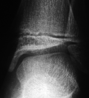
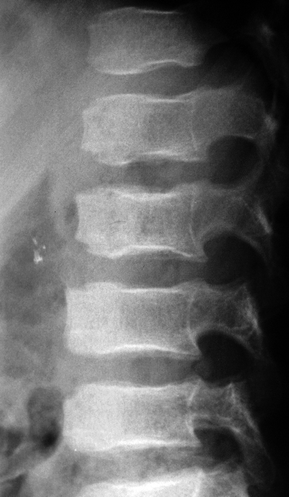
Fig. 8.6
Radiograph of the left ankle of a 15-year-old boy with SCD and tibiotalar slant. Even though this is a classic radiographic finding, other diseases, such as hemophilia and juvenile idiopathic arthritis, may present a similar appearance
Fig. 8.7
Lateral view of the thoracolumbar spine of a child with SCD showing the typical “H-shaped” vertebrae. There are focal depressions of the affected endplates, whose appearance differs from that of the biconcave vertebrae weakened by hyperplasia of the red marrow
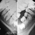
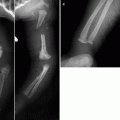
The so-called hand-foot syndrome (sickle-cell dactylitis) is characterized by recurrent episodes of pain and swelling of hands and feet related to infarction of the diaphyses of the small tubular bones. It is typical of small children (before age 2), consisting in one of the earliest manifestations of SCD. Radiographs are insensitive during the early stages, and characteristic radiographic findings (which include mottled osteoporosis, periosteal reaction, bone neoformation, and cortical thickening) may take some weeks to become apparent (Fig. 8.8). Moth-eaten lesions may be seen in more severe cases, and in long-standing disease, there is squaring and widening of the small tubular bones of hands and feet (Figs. 8.2 and 8.9).
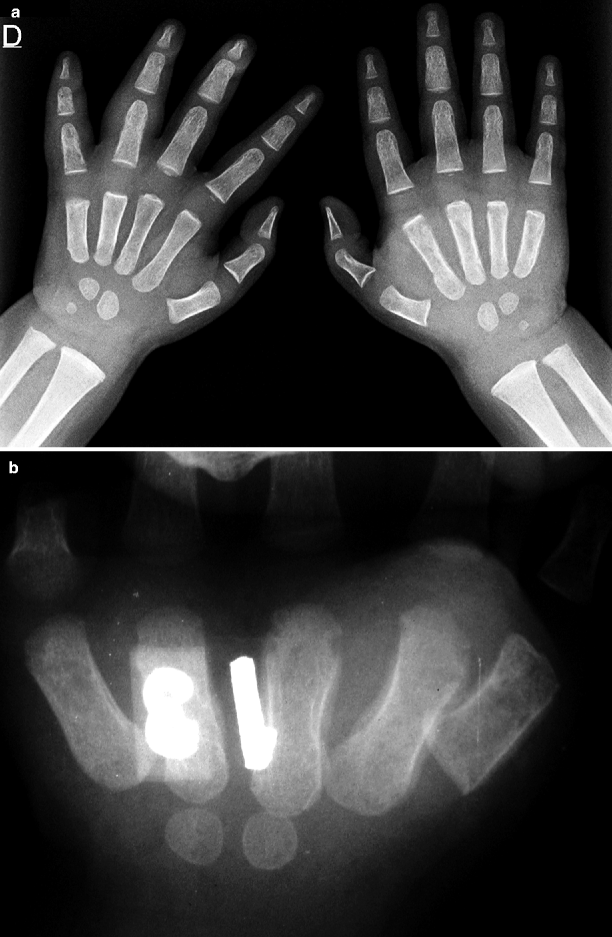
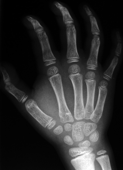
Fig. 8.8
In (a), radiograph of hands and wrists of a child with sickle-cell dactylitis demonstrates soft-tissue swelling and irregular periosteal reaction in the left metacarpals (from the second to the fifth) and in the right proximal phalanges of the fourth and fifth digits. There is also symmetric, diffuse, and bilateral enlargement of the phalanges, which show prominent bone trabeculae and solid periosteal reaction in some diaphyses. In (b), radiograph of the right hand of an 11-month-old male with acute dactylitis shows periosteal reaction in several metacarpals and cortical erosions, mainly in the third one; mottled osteoporosis is also present
Fig. 8.9
There is diaphyseal widening of the first metacarpal (which displays a cylindrical appearance in this child with SCD) due to expansion of the medullary cavity. Bone trabeculae are prominent and coarse, with a striated pattern. Solid periosteal reaction is seen in the proximal phalanges, mainly in the fourth finger
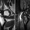
Painful episodes of vasoocclusion leading to acute bone infarction are the most frequent musculoskeletal complication of SCD after the second year of life, affecting mainly the long bones, vertebrae, and ribs. Even though radiographs are often normal in the early stages, lucent areas appear as bone destruction advances (Fig. 8.10). Chronic bone infarcts in the long bones appear as centrally located metadiaphyseal/diaphyseal lesions that exhibit a serpiginous calcified rim (Figs. 8.11 and 8.12), while marked sclerosis is found in chronic infarcts of the pelvic bones and of the vertebrae (Fig. 8.13). Additional findings that may be related to chronic infarcts include cortical thickening, layered periosteal reaction, and a “bone-within-bone” appearance (Figs. 8.11, 8.12, 8.13, and 8.14). Bone infarcts usually display normal or decreased uptake on bone scintigraphy in the first few days after the ischemic insult, showing increased uptake after the onset of revascularization; necrotic areas will remain “cold,” while ischemic bone with adequate revascularization will return to its normal appearance within a few months. On MRI, acute infarctions are ill-defined and may present a nonspecific edematous appearance; in time, however, hemoglobin degradation products eventually appear (showing high signal intensity on T1-WI and T2-WI), and heterogeneous post-gadolinium enhancement is often present (“ink stain” pattern), with surrounding bone marrow edema (Figs. 8.13, 8.15, and 8.16). On the other hand, chronic bone infarcts are geographic and well defined, presenting a hypointense rim on T1-WI and T2-WI and variable signal intensity in the central portion (Fig. 8.16). Avascular necrosis (epiphyseal infarct) in SCD is more common in adults than in children. The epiphyses of the proximal femur and of the proximal humerus are the preferential sites, with imaging findings similar to those of Legg-Calvé-Perthes disease, discussed in Chap. 7; lesions are usually larger and more frequently bilateral in SCD if compared to other causes of avascular necrosis (Figs. 8.13, 8.17, and 8.18). Subperiosteal fluid collections and hematomas are occasionally found in the limbs (Fig. 8.19), although they are more common in the orbits, associated with bone infarcts.
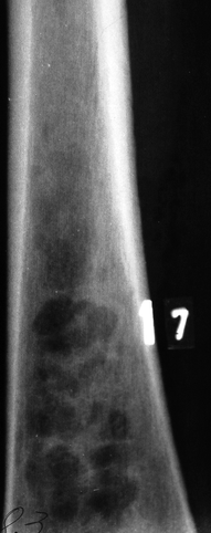
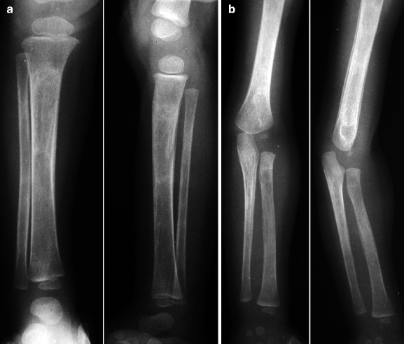
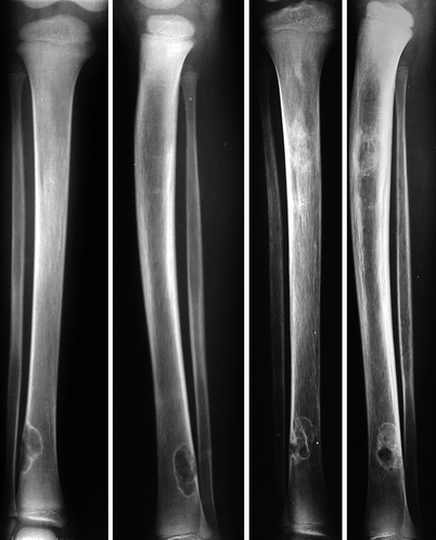
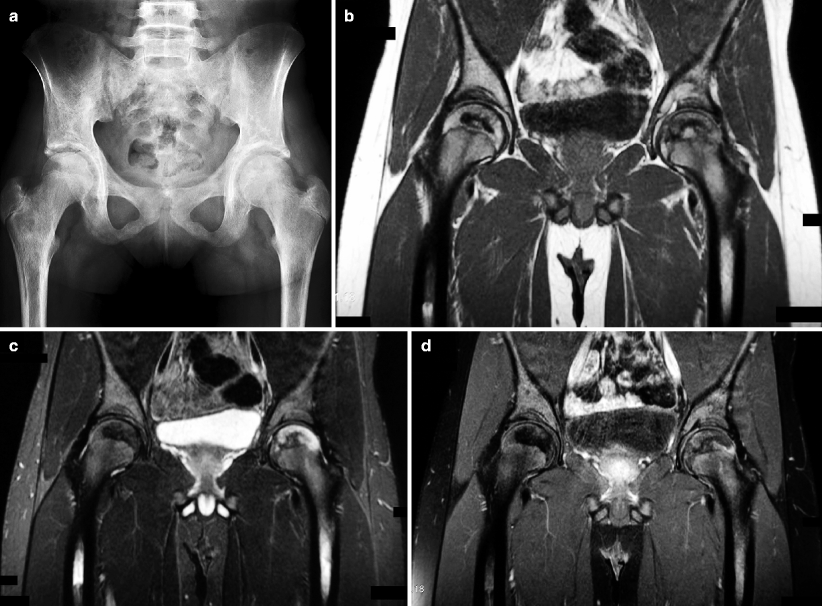
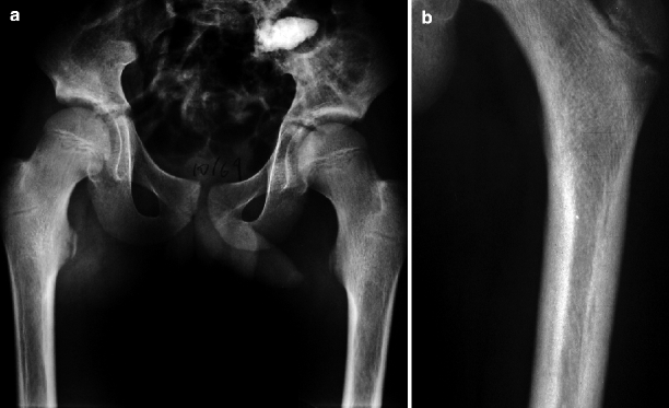

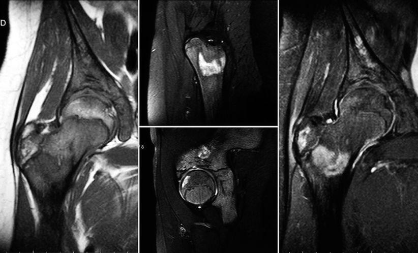
Fig. 8.10
Bone infarct of the right femur of a male adolescent with SCD. An extensive lucency with lobulated borders can be seen in the medullary cavity of the distal diaphysis, without associated calcifications. The distal portion of the lesion is reasonably well delimited, while the proximal portion is ill-defined
Fig. 8.11
Bone infarcts in a child with SCD (1 year and 6 months old). Radiographs of the right leg (a) and of the left upper limb (b) reveal diffuse osteoporosis, areas of bone sclerosis, and solid periosteal reaction in the long bones
Fig. 8.12
Anteroposterior and lateral views of the left leg of the same child, taken 3 years apart, demonstrating the low sensitivity of this method for the assessment of acute infarcts in SCD. At left, radiographs obtained during an acute painful crisis do not reveal abnormal findings, except for a non-ossifying fibroma in the distal tibia. The right images disclose sclerotic areas in the medullary cavity of the proximal tibia, with periosteal reaction and cortical thickening, representing chronic bone infarcts
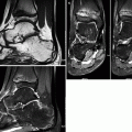
Fig. 8.13
A 16-year-old female with long-standing SCD. Pelvic radiograph (a) shows sclerotic areas in the medullary bone of the iliac bones and of the femora, with “bone-within-bone” appearance in the proximal femoral diaphyses. Even though it is clear that there are multiple chronic bone infarcts, this method is insensitive to detect acute ischemia. Coronal T1-WI (b), fat sat T2-WI (c), and post-gadolinium fat sat T1-WI (d) of the hips of the same patient reveal recent bone infarcts in the femoral diaphyses, with hyperintense areas on T1-WI and T2-WI and mild post-contrast enhancement. There is also extensive avascular necrosis of the left femoral head, with enhancement of the lateral pillar. A non-enhancing, serpiginous area of low signal intensity in all sequences in the right femoral head represents an old infarct, with bone sclerosis
Fig. 8.14
In (a and b), radiographs of distinct patients demonstrate linear areas of sclerosis in the medullary cavities of the proximal femora, adjacent to the cortical bone (“bone-within-bone”). In SCD, this finding – which may be found in many other conditions – is related to osteonecrosis
Fig. 8.15
An 8-year-old child with SCD and acute painful crisis in the right thigh. In (a), coronal fat sat T1-WI pre- (left) and post-gadolinium (right) reveal an extensive area of heterogeneous hyperintensity in the medullary cavity of the femur, representing bone infarct and the presence of hemoglobin degradation products. There is post-contrast enhancement of the ischemic bone and of the surrounding soft tissues. In (b) (transverse post-gadolinium fat sat T1-WI of the thighs taken 1 month later), the bone infarct of the right femur displays predominantly peripheral enhancement. Diaphyseal osteomyelitis in the contralateral femur is also present, with inflammation of the adjacent soft tissues, where diffuse and intense post-contrast enhancement can be seen. An intraosseous abscess is present in the left femur, appearing as an encapsulated fluid collection with subperiosteal extension and peripheral enhancement. Diaphyseal osteomyelitis of the left tibia was also present (not shown)
Fig. 8.16
 Legg-Calvé-Perthes Disease
 Peculiar Aspects of the Anatomy and Development of the Growing Skeleton
 Sports-Related Musculoskeletal Lesions in Pediatric Patients
 Juvenile Idiopathic Arthritis
 Juvenile Spondyloarthropathies and Pediatric Collagen Vascular Disorders
 Accidental and Non-accidental Articular Injuries in the Skeletally Immature Patient


T1-WI (left), fat sat T2-WI (center), and post-gadolinium fat sat T1-WI (right) of the right hip of a 15-year-old female with SCD. Geographic areas of abnormal signal intensity can be seen in the apophysis of the greater trochanter and in the supra-acetabular iliac bone, as well as in the femoral epiphysis and in the medullary cavity of the proximal diaphysis. The former two are heterogeneous lesions with fat in their central portion surrounded by a thin rim of low signal intensity in all sequences, while the latter two are ill-defined, presenting low signal intensity on T1-WI and high signal intensity on fat sat T2-WI; predominantly peripheral post-gadolinium enhancement is seen in all lesions. These are bone infarcts in different stages: the apophyseal and the iliac lesions are older, while the epiphyseal and diaphyseal ones show evidence of recent ischemia
Related posts:
Legg-Calvé-Perthes Disease
Peculiar Aspects of the Anatomy and Development of the Growing Skeleton
Sports-Related Musculoskeletal Lesions in Pediatric Patients
Juvenile Idiopathic Arthritis
Juvenile Spondyloarthropathies and Pediatric Collagen Vascular Disorders
Accidental and Non-accidental Articular Injuries in the Skeletally Immature Patient
Stay updated, free articles. Join our Telegram channel
Full access? Get Clinical Tree
