Musculoskeletal Ultrasound
US evaluation of the musculoskeletal system is a rapidly expanding area of sonographic diagnosis. Indications include assessment of soft tissue infections, detection and characterization of soft tissue masses, and the evaluation of muscle, tendon, and joint abnormalities [1,2,3]. This chapter highlights some of these areas of musculoskeletal sonography [4].
Imaging Technique
Most structures imaged are superficial; therefore, high-frequency (7-10 MHz) linear array transducers are most useful [4]. Interaction between the patient and the examining physician is essential to accurate examination. Consider the history and precise location of symptoms. Correlate the examination with point tenderness and location of pain with motion. Real-time observation during compression with the transducer yields vital additional information about the nature of visualized structures. Comparison with normal structures on the opposite side of the body may be extremely helpful. Color flow and spectral Doppler provide vital information about the vascularity of masses and inflammatory processes. Anisotropic artifact (see Chapter 1) is a prominent feature of US examination of tendons and ligaments. Because of their longitudinal fibrillar structure, tendons and ligaments appear echogenic when imaged perpendicular to the US beam and appear hypoechoic when imaged at an angle to the US beam [5].
Anatomy
Subcutaneous Fat
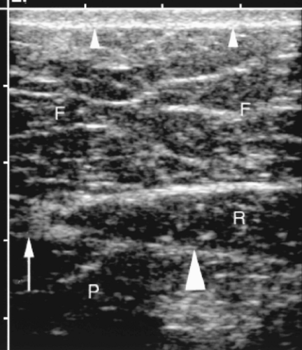
Subcutaneous fat is found just below the covering layer of skin.
Subcutaneous fat is hypoechoic and interspersed with thin linear septations of connective tissue. Thickness is related to the patient’s state of obesity (Fig. 13.1).
Skeletal Muscle
Skeletal muscle fibers are grouped into bundles defined by fibroadipose septa [4]. Dense connective tissue covers the entire muscle and additional thick fascia separates individual muscles.
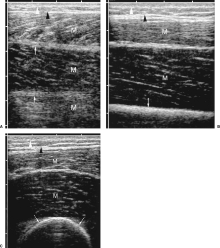
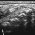
On longitudinal scans, muscles are hypoechoic with a pattern of fine echogenic strands in an oblique orientation. These strands correspond to fibroadipose septations (Fig. 13.2).
On transverse scans, the septa appear punctate or linear and are diffusely scattered over the hypoechoic background of muscle tissue (Fig. 13.2).
Doppler shows moderate blood flow within muscle tissue.
Dynamic imaging during contraction and relaxation illustrates function and aids in the detection of abnormalities.
Diffuse fat infiltration of muscle occurs with obesity and diffusely increases the echogenicity of muscle [6].
Tendons
Tendons in all areas of the body have a fairly uniform, clearly recognizable appearance.
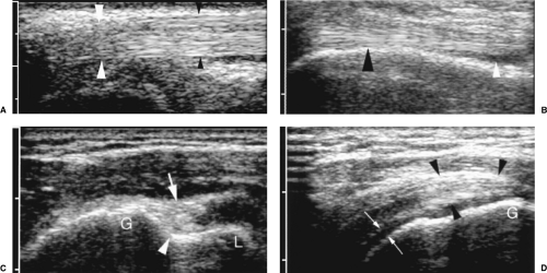
In longitudinal plane, tendons have a fine, linear, fibrillar internal pattern of parallel hyperechoic lines (Fig. 13.3). Higher frequency US shows more detail with finer and more numerous fibrillar echoes [7].
On transverse section, tendons are hyperechoic and round or oval in shape.
Synovial sheaths cover many tendons and appear as a thin (1-2 mm) hypoechoic rim surrounding the echogenic tendon (Fig. 13.3B). The long biceps tendon of the shoulder and the tendons of the wrist and ankle have synovial sheaths. The rotator cuff, Achilles, patellar, gastrocnemius, and semimembranous tendons do not have sheaths [4].
Tendons will routinely demonstrate the anisotropic effect (Fig. 13.3A) [5].
Ligaments
Ligaments attach bone to bone to provide stabilization.
Ligaments appear similar to tendons but have a more compact hyperechoic fibrillar pattern. Collagen fibers are most interweaved and more irregular in appearance than tendons.
Peripheral Nerves
Nerves appear as tubular, echogenic structures slightly less echogenic than tendons and ligaments. Multiple, parallel, linear internal echoes are characteristic.
Synovial Bursa
Bursa are potential spaces than normally contain only a tiny volume of fluid.
Normal bursa appear as flattened hypoechoic structures 1-4 mm in thickness in characteristic locations.
Bone Cortex
Because bone avidly absorbs sound energy, only the superficial surface of bone is evaluated by US.
Bone cortex appears as a bright echogenic surface with prominent posterior shadowing (Figs. 13.2B, C; 13.3C, D).
Hyaline Articular Cartilage
Hyaline cartilage covers the articular cortical surface of bone.
Cartilage is seen as a thin hypoechoic rim that covers the echogenic bone cortex (Fig. 13.3D).
Hyaline Epiphyseal Cartilage
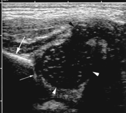
Sound transmits well through hyaline cartilage, allowing US evaluation of developing bone such as the infant hip [10,11].
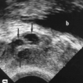
In the newborn infant the femoral head and trochanters consist entirely of cartilage. The hyaline cartilage appears diffusely hypoechoic with a pattern of echogenic dots scattered throughout its substance (Fig. 13.4). A pattern of echogenic vertical or spiral columns may also be seen. The nucleus of ossification appears as a highly echogenic focus in the center of the hypoechoic head. This focus progressively enlarges as ossification advances [12].
Musculoskeletal Masses
US can characterize the cystic or solid nature and vascularity of a mass but, with few exceptions, US cannot determine whether a solid mass is benign or malignant. US is effectively used to guide biopsy of soft tissue masses [13,14]. Soft tissue masses are described as to location, size, shape, margins, vascularity, deformability, and number of lesions.
Lipoma
Lipomas are a common mass of the superficial soft tissues. The tumor is benign and consists entirely of fat bounded by a thin capsule. The tumor may be lobulated in contour and divided by fibrous septa.
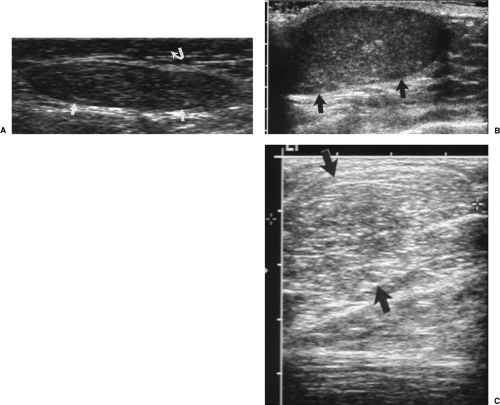
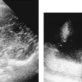
Knowing that fat in the abdomen is usually quite echogenic, it is somewhat surprising to recognize that subcutaneous lipomas appear moderately hypoechoic. Echogenicity is homogeneous. Lipomas may be isoechoic or mildly hyper- or hypoechoic compared to subcutaneous fat (Fig. 13.5) [15].
Lipomas appear well defined when surrounded by fibrous tissue or muscle but their margins are often indistinct when surrounded by fatty tissue.
Lipomas are oval in shape with the long axis of the tumor parallel to the skin [15].
Correlation with physical examination is useful to define the borders of the mass and to confirm the soft fluctuant nature of fat.
Tumor heterogeneity, large size, and prominent lobulations are signs that suggest malignancy (liposarcoma).
Hemangioma
Hemangioma is the second most common benign tumor of muscle and subcutaneous tissues. Hemangiomas consist of endothelial-lined vascular spaces of varying size with a variable amount of intervening fibrofatty tissue [16].
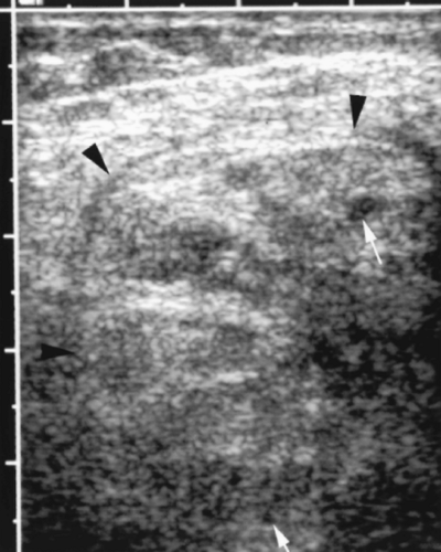
Hemangiomas are heterogeneous masses with tortuous blood vessels often visible coursing through the mass (Fig. 13.6).
Color flow Doppler shows prominent blood flow. A high vessel density (more than 5 vessels/cm2) and high flow velocity is characteristic [17].
Shadowing punctate echogenic foci represent phleboliths, which are characteristically present.
Nerve Tumors
Tumors arising from peripheral nerves are usually classified as schwannomas (also called neurinoma or neurilemoma) or neurofibromas. Malignant tumors are sarcomas that arise
from neurofibromas. Von Recklinghausen neurofibromatosis is characterized by widespread neurofibromas that present as skin nodules [9,18].
from neurofibromas. Von Recklinghausen neurofibromatosis is characterized by widespread neurofibromas that present as skin nodules [9,18].
Nerve tumors appear as well-defined, fusiform, hypoechoic masses. Lesions may be solitary, multiple, or elongated plexiform masses. A neurogenic origin is confirmed if careful scanning confirms that the soft tissue mass is connected to a nerve bundle at its proximal and distal poles [9].
Schwannomas tend to be more eccentric to the nerve and appear homogeneous with posterior acoustic enhancement. Doppler shows prominent vascularity. Cystic changes may occur.
Neurofibromas are more echogenic and coarse in appearance. Doppler shows low vascularity. A sonographic target appearance with peripheral low echogenicity surrounding central higher echogenicity has been described [19]. This sonographic finding corresponds to the MR target sign of high intensity peripherally and low intensity centrally seen on T2-weighted images.
US cannot reliably differentiate benign from malignant nerve sheath tumors. Findings that suggest malignancy include rapid growth, indistinct tumor margins, and invasion of adjacent tissue. Fine needle aspiration biopsy can be performed with US guidance. Needle insertion into the nerve causes intense pain.
Stay updated, free articles. Join our Telegram channel

Full access? Get Clinical Tree


