Myocardial Ischemia
Katherine R. Birchard
CLINICAL HISTORY
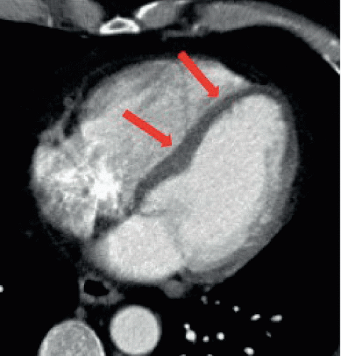
62-year-old female with chest pain, who underwent CTA to assess for aortic dissection.
FINDINGS
Figure 46A: Axial contrast-enhanced arterial-phase computed tomography angiography (CTA) image of the heart shows decreased attenuation of interventricular septum (arrows) of the left ventricle, compared with the lateral wall, which shows normal enhancement.
DIFFERENTIAL DIAGNOSIS
Myocardial ischemia, subendocardial infarct, artifact.
DIAGNOSIS
Myocardial ischemia.
Stay updated, free articles. Join our Telegram channel

Full access? Get Clinical Tree


