Chapter 10. Neck and Thyroid
Patient Preparation
• No preparation is required for neck and thyroid imaging.
Equipment and Technical Factors
• A high-frequency linear array is used for imaging the neck and thyroid.
• To image large superficial structure pathology, a curved linear or sector transducer with a stand-off pad to place the anatomy into the wider portion of the image may be used.
Imaging Protocol
• Longitudinal and transverse axes images of the organ or area of interest with the neck slightly extended (hyperextension is contraindicated).
• Demonstrate relational anatomy.
Variants
• Pyramidal lobe of the thyroid
Sonographic Measurements
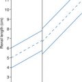
Thyroid
• Length: 4.0−6.0 cm
• Width: 1.3−1.8 cm
• Depth (AP): 1.3−1.8 cm
• Isthmus: 3.0 mm
Neck
• Length, depth, and width of cyst, mass, or diseased area.
| Neck and Thyroid | |||
|---|---|---|---|
| Sonographic Finding(s) | Clinical Presentation | Differential Diagnosis | Next Step |
Solid mass(es) more echogenic than thyroid with hypoechoic halo Well defined Possible cystic degeneration or “eggshell” calcification noted Predominately cystic mass(es) May appear complex | Asymptomatic Possible palpable nodule in thyroid | Thyroid adenoma Degenerating adenoma | Adenomas may undergo cystic degeneration and appear as complex mass Do not overlook the isthmus Use light transducer pressure to avoid missing small lesions |
| Cystic structure with irregular walls and internal echoes | Asymptomatic Possible palpable thyroid nodule | Thyroid cyst (with/without hemorrhage) Degenerating adenoma | Do not overlook the isthmus Use light transducer pressure to avoid missing small lesions |
Solitary hypoechoic mass with irregular border and microcalcifications Enlarged lymph node(s) in neck may be noted; the node(s) may be predominately cystic Lesion may be very small to large
Related posts:Stay updated, free articles. Join our Telegram channel
Full access? Get Clinical Tree


| |||