Fig. 1
(a, b) Surface anatomy of the convexity. (a) Sylvian fissure (dark area). The margins of the frontal, parietal, and temporal opercula are defined by the configuration of the sylvian fissure (S), its five major rami (the anterior horizontal ramus (AH), anterior ascending ramus (AA), posterior horizontal ramus (PHR), posterior ascending ramus (PA), and posterior descending ramus (PD), and its minor arms, the anterior subcentral sulcus (single arrowhead), posterior subcentral sulcus (double arrowheads), and transverse temporal sulci (triple arrowheads). (b) Cerebral convexity. The configuration of the sylvian fissure then permits identification of the adjoining gyri and sulci. GYRI: 1 superior frontal gyrus (SFG), 2 middle frontal gyrus (MFG), 3 inferior frontal gyrus (or, pars orbitalis; tr, pars triangularis; op, pars opercularis), 4 precentral gyrus (preCG), 5 postcentral gyrus (post CG), 6 supramarginal gyrus (SMG), 7 angular gyrus (AG), 8 superior temporal gyrus (STG), 9 middle temporal gyrus (MTG), 10 superior parietal lobule (SPL), 11 subcentral gyrus, 12 temporo-occipital arcus, 13 inferior temporal gyrus: * union of the MFG (2) with the preCG (4). SULCI; a superior frontal sulcus (SFS), b inferior frontal sulcus (IFS), c precentral sulcus (preCS), d central sulcus (CS), e postcentral sulcus (post CS), f superior temporal sulcus (STS), g intraparietal sulcus (IPS), h primary intermediate sulcus, j secondary intermediate sulcus, k accessory intermediate sulcus (not shown in this image), S sylvian fissure, single black arrowhead anterior subcentral sulcus, double black arrowheads posterior subcentral sulcus, triple black arrowheads transverse temporal sulci. Note specifically how the anterior horizontal and anterior ascending rami of the sylvian fissure divide the inferior frontal gyrus into the three partes orbitalis (or), triangularis (tr), and opercularis (op). The lateral orbital sulcus (l) separates the pars orbitalis from the lateral orbital gyrus (From Naidich et al. (1995); with permission)
2.1.2 Frontal Lobe
The convexity surface of the frontal lobe is formed by four gyri and three sulci (Fig. 2). The superior frontal gyrus (SFG) is a horizontally oriented, roughly rectangular bar of tissue that forms the uppermost margin of the frontal lobe. The middle frontal gyrus (MFG) is a horizontally oriented, undulant length of tissue that zigzags posteriorly to merge with the anterior face of the precentral gyrus. The inferior frontal gyrus (IFG) is a triangular gyrus that nestles inferiorly against the anteriormost portion of the sylvian fissure. The precentral gyrus (preCG) is a nearly vertical gyrus that forms the posterior border of the frontal lobe, behind the SFG, MFG, and IFG. The superior frontal sulcus (SFS) separates the SFG from the MFG. At its posterior end, the SFS bifurcates to form the superior precentral sulcus. The inferior frontal sulcus (IFS) separates the MFG from the IFG. At its posterior end, the IFS bifurcates to form the inferior precentral sulcus. Together, the superior and inferior portions of the preCS delimit the anterior face of the precentral gyrus, except where the MFG merges with the preCG between the superior and inferior precentral sulci.
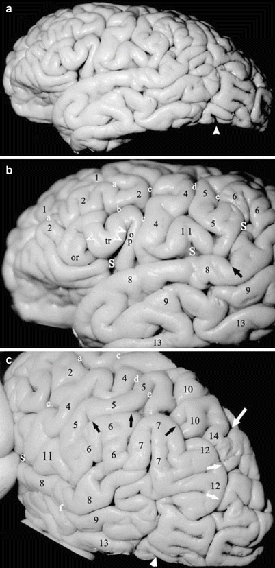
Fig. 2
(a–c) Normal cerebral convexity. (a) Anatomic specimen full surface. (b) Magnified view of the low-middle convexity. (c) Magnified oblique view of the parietal lobe. The surface vessels and the pia-arachnoid have been removed to expose the gyri and sulci more clearly (labels as in Fig. 1.). In (a), the single large white arrowhead = preoccipital notch. In (b), the portions of the sylvian fissure (S) are indicated: first white S = vallecula leading to the anterior horizontal ramus (single white arrow) and the anterior ascending ramus (double white arrow); second white S = posterior horizontal ramus; third white S = posterior ascending ramus; short black arrow = posterior descending ramus of the sylvian fissure. In (c), the anterior border of the occipital lobe extends from the lateral end of the parieto-occipital sulcus (large white arrow) above to the preoccipital notch (large white arrowhead) below. The intraparietal sulcus (multiple large black arrows) crosses the theoretical lobar border to become the intraoccipital sulcus (multiple large white arrows). The inferior parietal lobule is composed of the SMG (6), the AG (7), and the temporo-occipital arcus (second pli de passage of Gratiolet) (12). The superior parietal lobule merges into the superior occipital gyrus across the parieto-occipital border through the parieto-occipital arcus (first pli de passage of Gratiolet) (14) (a, b from Naidich et al. (1997); (c) From Naidich and Brightbill (1995); with permission)
The frontal gyri display characteristic configurations (Fig. 1) and variations (Albanese et al. 1989; Naidich et al. 1995, 1997). The IFG has an overall triangular configuration (hence, its synonym: triangular gyrus). The IFS courses above the IFG, bifurcates into the inferior preCS, and thereby separates the triangular IFG from the MFG above and from the preCG behind. The anterior horizontal and anterior ascending rami of the sylvian fissure extend upward into the triangular IFG, partially subdividing it into three portions: the pars orbitalis, which abuts the orbital gyri of the frontal lobe; the pars triangularis in the center; and the pars opercularis, which forms the anteriormost portion of the frontal operculum. Together, the three parts of the IFG resemble an oblique letter “M” (Figs. 1, 2, 3, and 4). Because the anterior ascending ramus of the sylvian fissure cuts through the full thickness of the IFG to reach the insula, the cortex of the pars opercularis presents both a superficial cortex visible on the surface and a deep cortex within the depths of the fissure (Naidich et al. 2001a).
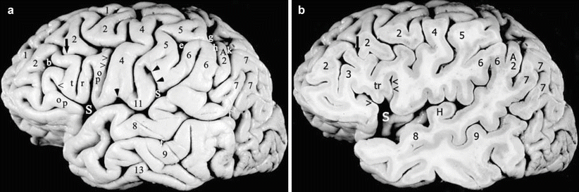
Fig. 3
(a, b) Normal variations in convexity anatomy. (a) Convexity surface of a prepared left hemisphere. (b) Sagittal section of a prepared left hemisphere. A vertically oriented connecting gyrus (short black arrow) crosses the inferior frontal sulcus (IFS) (b) to unite the pars triangularis (tr) with the middle frontal gyrus (MFG) (2). A sulcus triangularis (between the t and the r) deeply notches the superior surface of the pars triangularis making it appear bifid. The precentral gyrus (4) and the postcentral gyrus (5) unite beneath the central sulcus to form the subcentral gyrus (11), which is delimited by a shallow anterior subcentral sulcus (single black arrowhead) and a deep posterior subcentral sulcus (double black arrowheads). The inferior portion of the postcentral sulcus (e) forms the initial upswing of the arcuate intraparietal sulcus (IPS) (g). The posterior ascending ramus of the sylvian fissure (S) indents the inferior parietal lobule (IPL) to form the supramarginal gyrus (SMG) (6). The superior temporal sulcus (STS) (ƒ) indents the posterior portion of the IPL to form the angular gyrus (AG) (7). The entire STS parallels the sylvian fissure, hence, its synonym: parallel sulcus. The distal end of the STS within the AG may be designated the angular sulcus. In this specimen, an intercalated accessory preangular gyrus (A2) separates the SMG from the AG. Note the relationships of the IPS (g), the SMG (6), the AG (7), and the accessory preangular gyrus (A2) with the primary intermediate sulcus (h), the secondary intermediate sulcus (j), and the accessory intermediate sulcus (k). In (b), the sagittal section exposes the characteristic appearance of the transverse temporal gyrus of Heschl (H) on the superior surface of the temporal lobe (From Naidich et al. (1995); with permission)
2.1.3 Temporal Lobe
The convexity surface of the temporal lobe is formed by three horizontal gyri: the superior temporal gyrus (STG), the middle temporal gyrus (MTG), and the inferior temporal gyrus (ITG), separated by the superior temporal sulcus (STS) and the inferior temporal sulcus (ITS) (Fig. 5). The superior and middle temporal gyri extend posteriorly and then swing upward to join with the parietal lobe. The inferior temporal gyrus forms the inferior edge of the convexity surface of temporal lobe and curves onto the inferior surface of the temporal lobe. It is delimited posteriorly by a small notch, the preoccipital notch (synonyms: temporo-occipital notch or incisura), which separates the inferior temporal gyrus anteriorly from the inferior occipital gyrus posteriorly. The STS courses parallel to the posterior horizontal and the posterior ascending rami of the sylvian fissure (hence, its synonym, parallel sulcus). The posterior portion of the STS is directed superiorly and is sometimes designated the angular sulcus. The ITS courses approximately parallel to the inferior margin of the convexity and may become continuous posteriorly with the inferior occipital sulcus.
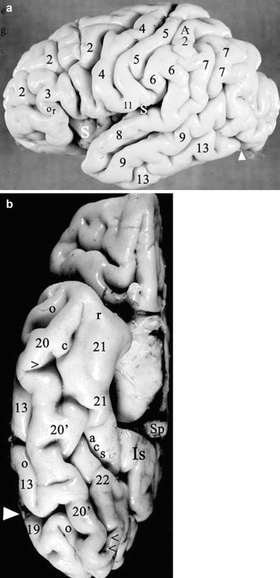
Fig. 5
(a, b) The temporal lobe. Prepared anatomic specimen of the right hemisphere from a 1-day-old girl. (a) Convexity (reversed to match the other lateral views). The simplified gyral and sulcal pattern reflects the young age. The inferior temporal gyrus (13) extends posteriorly to the preoccipital notch (large white arrowhead). An accessory preangular gyrus (A2) is situated superior to the SMG (6) and anterior to the AG (7). Labels as in Figs. 1 and 2. (b) Inferior surface. The brainstem and the inferior thalamus have been removed to reveal the medial surface more clearly. The inferior temporal gyrus (13) and the inferior occipital gyrus (19) form the inferior margin of the hemisphere, separated by the preoccipital notch (large white arrowhead). The lateral occipitotemporal sulcus (o) delineates their medial border and separates them from the lateral occipitotemporal gyrus (LOTG) (20) further medially. The medial occipitotemporal sulcus (synonym: collateral sulcus) (c) delimits the medial border of the LOTG over its full length. Anteriorly, the collateral sulcus approximates the rhinal sulcus (r), and may run into it, or may parallel it. The parahippocampal gyrus (PHG) (21) forms the medial surface of the temporal lobe over its full length and extends posteriorly to become the isthmus (Is) of the cingulate gyrus inferior to the splenium (Sp). In the anterior half of the temporal lobe, the collateral sulcus (c) separates the LOTG (20) from the PHG (21). In the posterior half, the medial occipitotemporal gyrus (MOTG) (lingual gyrus) (22) intercalates itself between the LOTG and the PHG. The collateral sulcus stays with the medial border of the LOTG, so the collateral sulcus (c) separates the LOTG (20) from the MOTG (22), while the anterior calcarine sulcus (acs) separates the MOTG (22) from the PHG (21). The anterior and posterior transverse collateral sulci (single and double black arrowheads) delimit a midportion of the LOTG (20 1 ) that has been designated by Duvernoy (Duvernoy 1991) as the fusiform gyrus (From Daniels et al. (1987); with permission)
2.1.4 Parietal Lobe
The convexity surface of the parietal lobe is formed by three portions: the vertically oriented postcentral gyrus (post CG) anteriorly, and the two superior parietal (SPL) and inferior parietal (IPL) lobules posteriorly (Figs. 1, 2, 3, and 4). The postCG courses vertically just posterior and parallel to the preCG. The postCG is separated from the preCG by the intervening central sulcus (CS) over most of its length. However, inferiorly, the postCG merges with the preCG inferior to the CS along the subcentral gyrus (subCG) just above the sylvian fissure. Superiorly, the postCG merges with the preCG superior the CS along the paracentral lobule (paraCL) on the medial surface of the hemisphere. Thus, the precentral and postcentral gyri actually form a continuous band of tissue that circles around the central sulcus from the precentral gyrus through the subcentral gyrus, into the postcentral gyrus and the paracentral lobule, returning into the precentral gyrus.
The posterior border of the post CG is delimited by the superior and inferior postcentral sulci. The superior postCS separates the upper postCG from the superior parietal lobule. The inferior postCS separates the postCG from the inferior parietal lobule. The inferior postCS may be considered the upswing of a long, deep, arcuate intraparietal sulcus (IPS) that ascends behind the lower postCG and then slashes posteriorly across the convexity surface of the parietal lobe, dividing it into the SPL situated superomedial to the IPS and the IPL situated inferolateral to the IPS. The posterior downswing of the arcuate IPS then crosses the theoretical border between the parietal and occipital lobes and continues into the occipital lobe, where it is designated the intraoccipital sulcus (IOS) (synonym: superior occipital sulcus, SOS) (Figs. 1, 2, 3, and 4). Posteriorly, the SPL becomes continuous with the superior occipital gyrus (SOG) behind it through a narrow band of tissue designated the arcus parietooccipitalis (first pli de passage of Gratiolet) (Fig. 2) (Duvernoy 1991).
Within the inferior parietal lobule, the posterior ascending ramus of the sylvian fissure swings upward into the anterior portion of the IPL and is capped by a horseshoe-shaped gyrus designated the supramarginal gyrus (SMG). In parallel fashion, the distal STS swings upward into the posterior portion of the IPL where it is capped by a second horseshoe-shaped gyrus designated the angular gyrus (AG). The AG usually lies just posterior to the SMG, but may be displaced from that position by variant accessory gyri (Figs. 1, 2, 3, and 4) (Naidich et al. 1995, 1997). Together the SMG and the AG constitute most of the IPL. An additional small horseshoe of tissue, designated the second parieto-occipital arcus (second pli de passage of Gratiolet), connects the AG with the middle OG posterior to it, completing the IPL (Fig. 2) (Duvernoy 1991). A small primary intermediate sulcus descends from the IPS to separate the SMG from the AG. A small secondary intermediate sulcus descends from the IPS to define the posterior border of the AG, separating the AG from the rest of the IPL posterior to it (Naidich et al. 2001a).
2.1.5 Occipital Lobe
The convexity surface of the occipital lobe has also been divided into three horizontal gyri: the superior occipital gyrus (SOG), the middle occipital gyrus (MOG), and the inferior occipital gyrus (IOG). These are separated by the superior and inferior occipital sulci (SOS and inferior OS). The SOS (synonym: intraoccipital sulcus) is the direct continuation of the IPS (Figs. 1, 2, 3, and 4). The inferior OS is usually coextensive with the inferior temporal sulcus. Therefore, the MOG lies just posterior to the confluence of the temporal and parietal lobes at the SMG, the AG, the STG, and the MTG. The MOG is the largest portion of the occipital lobe on the convexity. It is usually subdivided into a superior and an inferior portion by a horizontally oriented middle OS (synonym: lateral OS).
2.1.6 Insula
Separating the superior and inferior covers (opercula) of the sylvian fissure discloses the Island of Reil (insula). The insula is delimited circumferentially by the peri-insular sulcus (synonym: circular sulcus), composed of the anterior, superior, and inferior perisylvian sulci (Ture et al. 1999). The central sulcus of the convexity extends over the insula as the central sulcus of the insula, dividing the insula into a larger anterior and a smaller posterior lobule (Fig. 6) (Naidich et al. 2004; Nieuwenhuys et al. 1988; Ture et al. 1999). The anterior lobule typically has three vertically oriented short insular gyri designated the anterior short, middle short, and posterior short insular gyri. These three converge anteroinferolaterally to form the apex of the insula. The posterior lobule of the insula typically displays two oblique gyri: the anterior and posterior long insular gyri. The anterior insula is connected exclusively to the frontal lobe, whereas the posterior insula is connected to both the temporal and the parietal lobes (Naidich et al. 2004; Ture et al. 1999).
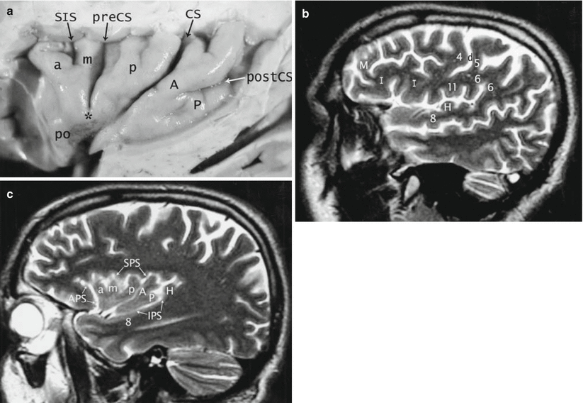
Fig. 6
(a–c) Insula. (a) Prepared anatomic specimen of the convexity surface of the insula, after removal of the overhanging opercula. A 71-year-old man. The central sulcus (CS) extends across the triangular insula like a “hockey stick,” dividing it into a larger anterior lobule and a smaller posterior lobule. The anterior lobule typically displays three gyri, the anterior short (a), middle short (m), and posterior short (p) insular gyri, separated by the short insular sulcus (SIS) and the precentral sulcus (preCS). These three characteristically converge inferiorly to form the apex of the insula. The posterior insular lobule typically displays two gyri, the anterior long (A) and the posterior long (P) insular gyri, separated by the postcentral sulcus (postCS). These too often merge together, anteriorly, just behind the central sulcus. Just inferomedial to the apex , the pole of the insula (po) forms the most anteroinferomedial portion of the insula. The central sulcus courses under the apex and the pole and then abruptly swings medially toward the suprasellar cistern. (b, c) Normal sagittal T2-weighted MRI. A 51-year-old man. Laterally (b), the convexity gyri form two opercula that cover the insula. Superiorly, the inferior frontal gyrus (I), the inferior ends of the precentral (4) and postcentral (5) gyri, the subcentral gyrus (11), and the supramarginal gyrus (6) form the frontoparietal operculum. The superior temporal gyrus (8) and Heschl’s gyrus (H) form the temporal operculum. M middle frontal gyrus. Medially (c), the insula is delimited by the peri-insular sulcus, composed of three segments: the anterior (APS), superior (SPS), and inferior (IPS) peri-insular sulci. The anterior (a), middle (m), and posterior (p) short insular gyri constitute the larger anterior lobule, while the anterior (A) and posterior (P) long insular gyri form the smaller posterior lobule. The central sulcus courses between the two lobules. Heschl’s gyrus (H) snugs up against the posteromedial portion of the posterior long insular gyrus. 8 = superior temporal gyrus (From Naidich et al. (2004) with permission)
2.2 Inferior Surface
2.2.1 The Frontal Lobe
The inferior (orbital) surface of the frontal lobe is formed by the gyrus rectus medially and four orbital gyri laterally, separated by the olfactory and orbital sulci. The gyrus rectus forms the medial margin of the orbital surface of frontal lobe for the full length of the frontal lobe. The lateral border of the gyrus rectus is delimited by the olfactory sulcus (Fig. 7). The orbital gyri are arranged around an “H-shaped” orbital sulcus as the medial orbital, lateral orbital, anterior orbital, and posterior orbital gyri.
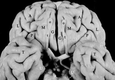
Fig. 7
Inferior surface of frontal lobe. Prepared anatomic specimen of the inferior surfaces of the anterior brain. The inferior surface of the frontal lobes is divided into two portions. The paired paramedian linear gyri recti (G) flank the interhemispheric fissure and are delimited laterally by the olfactory sulci, largely obscured here by the olfactory bulbs (o) and tracts (t). Lateral to the olfactory sulci, the orbital gyri of the frontal lobe are arranged around approximately H-shaped orbital sulci as the paired medial (M), lateral (L), anterior (A), and posterior (P) orbital gyri. In true base view, the anterior temporal lobes (T) overlap the posterior orbital gyri. Note the paired unci (U)
2.2.2 The Temporo-occipital Lobes
The inferior surface of the temporo-occipital lobe is formed by the inferior temporal gyrus, the lateral occipitotemporal gyrus (LOTG), the medial occipitotemporal gyrus (MOTG) (synonym: lingual gyrus), and the parahippocampal gyrus (PHG), separated by the lateral occipitotemporal sulcus, the collateral sulcus, and the anterior calcarine sulcus (Fig. 5). Medially, the parahippocampal gyrus forms the medial border of the temporal lobe for the full length of the temporal lobe, from just posterior to the temporal pole to the level of the splenium. Posterior to the splenium, the medial occipitotemporal gyrus forms the medial border of the occipital lobe. Laterally, along the full length of the temporo-occipital lobes, the inferior temporal gyrus and the inferior occipital gyrus curve medially, form the inferior cerebral margin, and pass onto the inferior surface, where they constitute the lateralmost portion of the inferior surface of the temporo-occipital lobes. Only the small preoccipital notch delimits the inferior temporal gyrus from the inferior occipital gyrus. Centrally, the LOTG runs the full length of the temporo-occipital lobes from the temporal pole to the occipital pole. Throughout its length, the LOTG remains just medial to the ITG and the IOG and is separated from them by the lateral occipitotemporal sulcus. Medially, the medial occipitotemporal sulcus (synonym: collateral sulcus) runs the full length of the LOTG (Fig. 5b). In the anterior half of the temporal lobe, the lateral occipitotemporal sulcus separates the LOTG from the parahippocampal gyrus. In the posterior half of the temporal lobe, the medial occipitotemporal gyrus intercalates itself between the LOTG and the parahippocampal gyrus. Therefore, in the posterior half of the temporal lobe, the lateral occipitotemporal sulcus separates the LOTG form the MOTG, while the anterior calcarine sulcus separates the MOTG from the PHG.
Two synonyms are commonly used for temporo-occipital gyri. The term lingual gyrus usually refers to the MOTG (Duvernoy 1991; Ono et al. 1990). The term fusiform gyrus is most often used to designate either the LOTG (Ono et al. 1990) or a large middle portion of the LOTG that crosses the arbitrary border of the temporal and the occipital lobes between the anterior and the posterior transverse collateral sulci (Duvernoy 1991). However, in another usage, Williams et al. (1989) group the MOTG and the LOTG together as the fusiform gyrus. In most brains, the fusiform gyrus is larger on the left (Kopp et al. 1977; Naidich et al. 2001a).
2.3 Superior Surface of the Temporal Lobe
Opening the margins of the sylvian fissure, or resecting the overlying superior operculum, displays the superior surface of the temporal lobe, designated the superior temporal plane. The prominent features of this plane are one or more transverse temporal gyri (of Heschl) (Fig. 8). Heschl’s gyrus or gyri (HG) arise just posteromedial to the insula and course obliquely across the superior temporal surface from posteromedial to anterolateral and may be visible at the external surface of the sylvian fissure. The presence of an HG is constant. The number of HG on each side and their symmetry are highly variable. Heschl’s gyri may be single (66–75 %), double (25–33 %), or triple (1 %), both unilaterally and bilaterally (Yoshiura et al. 2000; Yousry et al. 1997a). Heschl’s gyrus is often larger and longer on the left side than the right, but there is no constant relationship between HG and the side of handedness or cerebral dominance (Carpenter and Sutin 1983; Yousry et al. 1997a). A shallow longitudinal sulcus (of Beck) may groove the superior surface of HG, especially laterally, giving it a partially bifid appearance. A deep transverse temporal sulcus (Heschl’s sulcus [HS]) typically defines the posterior border of HG.
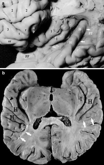
Fig. 8
(a, b). The superior surface of the temporal lobe from prepared anatomic specimens. (a) Anatomy of Heschl’s gyrus in relation to the insula. Resection of the frontal and parietal opercula exposes the middle frontal gyrus (2); the pars orbitalis (or) triangularis (tr) and the residual portion of the opercularis (op) of the inferior frontal gyrus (3); the oblique long gyri (black I) of the posterior insula; the length of a single Heschl’s gyrus (H) which courses obliquely across the superior surface of the temporal lobe from just posterior to the insula (posteromedially) toward the convexity surface of the superior temporal gyrus (anterolaterally); Heschl’s sulcus (white arrows) immediately posterior to Heschl’s gyrus; the planum polare (PP) anterior to the HG; and the planum temporale (PT) posterior to HG (From Naidich and Matthews (2000) with permission). (b) Exposure of the superior surfaces of both temporal lobes by resection of the overlying frontal and parietal opercula. Right is to the reader’s right. The single right and the dual left Heschl’s gyri (H) course obliquely across the upper surface of the temporal lobe from just posterior to the insulae (I) (posteromedially) to the convexity surface (anterolaterally). A shallow Beck’s sulcus (small black arrowhead) grooves the left HG laterally. Heschl’s sulcus delimits the posterior border of HG. The planum polare extends from the temporal pole to the anterior aspect of HG. The planum temporale extends between Heschl’s sulcus and the posterior limit of the sylvian fissure (white arrows) and is substantially larger on the left
The oblique Heschl’s gyrus divides the superior surface of the temporal lobe into three parts. (1) The flat superior surface of the temporal lobe anterior to HG is designated the planum polare. (2) From HS at the posterior border of HG to the posterior end of the sylvian fissure, the flat superior surface of the temporal lobe is designated the planum temporale. (3) The posterosuperior extension of the planum temporale along the posterior bank of the posterior ascending ramus of the sylvian fissure may be designated the planum parietale. The planum temporale is triangular. It is typically asymmetric on the two sides and most often is larger on the left in humans, chimpanzees and other great apes (Fig. 8) (Gannon et al. 1998). The size of the planum temporale seems to correlate with the side of language dominance (and perhaps with right or left-handedness, gender, or both (Galaburda and Sanides 1980; Galaburda et al. 1998; Geschwind 1965a, b, 1970; Geschwind and Levitsky 1968; Pieniadz and Naeser 1984; Steinmetz and Seitz 1991; Steinmetz et al. 1989b, 1990a, b, 1991). Most of the variation in the sizes of the planum temporale may be ascribed to differing sizes of a cytoarchitectonic zone designated area Tpt (Galaburda and Sanides 1980). Area Tpt has a homologue in nonhuman primates that shows significant asymmetry (left > right) at the cellular level (Gannon et al. 1998).
2.4 Medial Surface
The medial surface of the cerebrum is arranged as a radial array of gyri and sulci that are oriented either co-curvilinear with the corpus callosum or perpendicular to it (Fig. 9). The major gyri of the medial surface are the cingulate gyrus (CingG), the superior frontal gyrus (SFG) (whose medial surface may also be designated the medial frontal gyrus), the paracentral lobule (ParaCL), the precuneus (PreCu), the cuneus (Cu), and the medial occipitotemporal gyrus (MOTG) (synonym: lingual gyrus). The major sulci are the callosal sulcus (CalS), the cingulate sulcus (CingS), the paracentral sulcus (ParaCS), the subparietal sulcus (SubPS), the parieto-occipital sulcus (POS), the calcarine sulcus (CaS), and the anterior calcarine sulcus (AntCaS). The cingulate gyrus encircles the corpus callosum. It is delimited from the corpus callosum centrally by the callosal sulcus and from the superior frontal gyrus and paracentral lobule superficially by the cingulate sulcus.
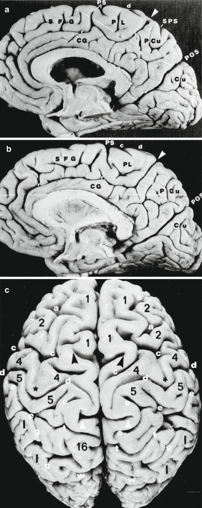
Fig. 9
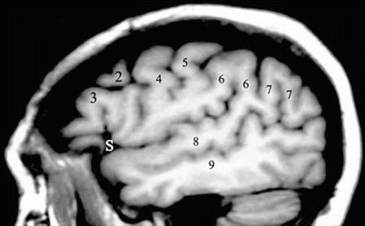
(a–c) Medial and superior surfaces of both hemispheres. Prepared anatomic specimens. (a, b) Medial surfaces. The major sulci subdivide the medial surface of each cerebral hemisphere into the cingulate gyrus (CG), medial surface of the superior frontal gyrus (SFG), paracentral lobule (PL), precuneus (PCu), and cuneus (Cu). The posterior end of the cingulate sulcus sweeps sharply upward toward the superior cerebral margin as the marginal portion of the cingulate sulcus (pars marginalis, white arrowhead), which separates the paracentral lobule anteriorly from the precuneus posteriorly. Anterior to the pars marginalis, the paracentral sulcus (PS) arises from the cingulate sulcus and/or from the cerebral margin to separate the superior frontal gyrus from the paracentral lobule. The vertical and horizontal arms of the “H-shaped” subparietal sulcus (s) groove the medial surface of the precuneus and delimit it from the cingulate gyrus inferior to it. The superior ends of the vertical arms of the subparietal sulcus may reach to and notch the superior margin. The superior medial end of the central sulcus (d) usually crosses over the cerebral margin onto the medial surface and then recurves sharply posteriorly to course nearly perpendicular to the pars marginalis, just millimeters in front of the pars marginalis. As a consequence, the most superior medial portion of the precentral gyrus (4) merges with the most superior medial portion of the postcentral gyrus (5) around the uppermost end of the central sulcus to form the paracentral lobule anterior to the pars marginalis. A posterior portion of the postcentral gyrus passes posterior to the pars marginalis to merge with the precuneus. The prominent parieto-occipital sulcus (POS) courses approximately parallel to the pars marginalis, but posterior to the splenium, and delimits the parietal lobe plus cingulate gyrus anterosuperiorly from the occipital plus temporal lobes postero-inferiorly. (c) Superior surface of the two hemispheres. The two cerebral hemispheres border the interhemispheric fissure (IHF). Multiple sulci oriented at right angles to the IHF form a series of transverse grooves or “crossbars” across the IHF. The paired partes marginales (white arrowheads) are often the most prominent of these grooves and extend laterally into the hemispheres for a substantial distance. At the vertex, the lateral edges of the two partes marginales often curve anteriorly to form a bracket, open anteriorly. The paired central sulci (d) undulate across the cerebral convexity between the precentral gyri (4) and the postcentral gyri (5). They typically hook sharply posteriorly as they circumscribe the hand motor region of the precentral gyrus , and then reverse curvature, become concave posteriorly, and converge toward the IHF just anterior to the partes marginales. The two central sulci (d) characteristically (but not invariably) pass anterior to and medial to the lateral edges of the partes marginales (“enter the bracket”) and reach to or cross the superior margins of the hemispheres. The postcentral gyri course superiorly, behind the precentral gyri, toward or to the cerebral margins. The medial ends of the postcentral sulci (e) usually bifurcate around the partes marginales to form a prominent “parenthesis” configuration (e, e in the right hemisphere). The interlocking curves of the precentral gyri (d), postcentral gyri (e), and the partes marginales (white arrowheads) form a characteristic set of interlocking curves that usually identifies these sulci and the adjacent gyri. Other labels: (1) superior frontal gyrus, (2) middle frontal gyrus, (4) precentral gyrus, (5) postcentral gyrus, (16) precuneus, (a) superior frontal sulcus, (c) precentral sulcus, (d) central sulcus, (e) postcentral sulcus, (g) intraparietal sulcus, (r) cingulate sulcus, (s) subparietal sulcus, (t) superior parietal sulcus, and (w) parieto-occipital sulcus (From Naidich et al. (1996); with permission)
The gyri that form the medial surface of the brain peripheral to the cingulate gyrus and sulcus are nothing more than the medial aspects of the gyri that constitute the high convexity of the brain. The superior frontal gyrus of the convexity curves over the cerebral margin onto the medial surface to form a broad arc of tissue designated the SFG or medial frontal gyrus. The precentral gyrus and the postcentral gyrus curve over the cerebral margin from the convexity onto the medial surface and join together to form the paracentral lobule. The superior parietal lobule curves over the cerebral margin onto the medial surface to form the precuneus. The superior occipital gyrus curves over the cerebral margin onto the medial surface to form the cuneus. The SFG is separated from the paracentral lobule posterior to it by the paracentral sulcus. The posterior end of the cingulate sulcus sweeps upward to reach the cerebral margin. This radially oriented distal portion of the cingulate sulcus is the pars marginalis (pM). The pars marginalis separates the paracentral lobule anteriorly from the precuneus posteriorly. The upper end of the central sulcus typically curves over the margin onto the medial surface of the hemisphere just anterior to the pars marginalis. This medial portion of the CS courses posteriorly nearly perpendicular to the pars marginalis. The “H”-shaped subparietal sulcus lies posterior to the pars marginalis and separates the inferior end of the precuneus from the cingulate gyrus deep to it. The parieto-occipital sulcus courses parallel to the pars marginalis, joins with the anterior end of the calcarine sulcus, and continues anteriorly as the anterior calcarine sulcus. The POS separates the precuneus anteriorly from the cuneus posteriorly. The calcarine sulcus separates the cuneus superiorly from the MOTG (lingual gyrus) inferiorly. The anterior calcarine sulcus separates the cingulate gyrus anteriorly from the MOTG posteriorly. The calcarine sulcus may remain entirely on the medial surface of the hemisphere, extend posteriorly to reach the occipital pole, or extend beyond medial surface onto either the convexity or inferior surfaces of the occipital lobe.
3 Lobar Borders
The precise borders of the temporal, parietal, and occipital lobes on the convexity are highly arbitrary. Published diagrams from different authors indicate substantially different criteria for partitioning the TPO lobes along the convexity (Yousry 1998). The very definitions of these borders and their lobes have evolved substantially over the years (Yousry 1998). In one common system of nomenclature, one identifies the lobes by first finding the lateral end of the deep parieto-occipital sulcus near the superior margin of the hemisphere. Then one identifies the inconstant preoccipital notch (Gusmão et al. 2002) in the inferior margin of the hemisphere (Fig. 10). The arbitrary anterior border of the occipital lobe is then defined by the drawing the “lateral parietotemporal line” along the convexity from the lateral end of the parieto-occipital sulcus above to the preoccipital notch inferiorly.
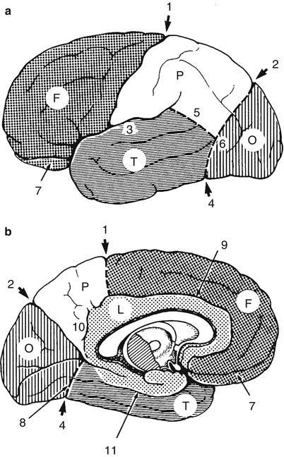
Fig. 10
(a, b) Lobar borders according to Ono, Kubik, and Abernathy. (a) Convexity. (b) Medial surface. The anterior border of the occipital lobe is delimited by the lateral parietotemporal line (6) drawn between the parieto-occipital sulcus and the preoccipital notch. On the convexity, the temporal lobe is separated from the parietal lobe by the temporo-occipital line (5) drawn from the sylvian fissure to the middle of the anterior border of the occipital lobe. On the basal surface, the temporal lobe is separated from the occipital lobe by the basal parietotemporal line (8) drawn from the preoccipital notch to the union of the parieto-occipital sulcus with the calcarine sulcus (unlabelled). F frontal lobe, O occipital lobe, P parietal lobe, T temporal lobe, 1 central sulcus, 2 parieto-occipital sulcus, 3 sylvian fissure, 4 preoccipital notch, 5 temporo-occipital line, 6 lateral parietotemporal line, 7 orbital surface, 8 basal parietotemporal line, 9 cingulate sulcus, 10 subparietal sulcus, 11 collateral sulcus (From Ono et al. (1990), p 9; with permission)
Next one demarcates the borders of the temporal and parietal lobes that abut onto the occipital lobe. To do this, one draws the “temporo-occipital line,” defined variably as (1) an arc from the distal end of the posterior descending ramus of the sylvian fissure to the midpoint of the anterior border of the occipital lobe (Ono et al. 1990), (2) an arc from the posterior descending ramus of the sylvian fissure to the anterior border of the occipital lobe, taking care to make sure that the arc is co-curvilinear with the IPS above (Duvernoy 1991; Schwalbe 1881; Yousry 1998), (3) an arc from the posterior descending ramus of the sylvian fissure to the anterior border of the occipital lobe at the preoccipital notch (Jensen 1871; Yousry 1998), or (4) a straight linear extension from the distal end of the posterior horizontal ramus of the sylvian fissure to the anterior border of the occipital lobe (Dejerine 1895; Talairach and Tournoux 1988; Yousry 1998).
On the medial surface, the deep parieto-occipital sulcus clearly divides the parietal lobe from the occipital lobe and is the landmark used to define the anterior border of the medial occipital lobe in all systems of nomenclature. On the inferior surface, the demarcation of the lobes again becomes arbitrary. On the inferomedial surface, one may delineate the temporal lobe from the occipital lobe by drawing a basal parietotemporal line from the preoccipital notch to, variably, (1) the inferior end of the parieto-occipital sulcus where it joins the calcarine sulcus (Fig. 10) (Ono et al. 1990), (2) the anterior calcarine sulcus beneath the splenium (Fig. 11) (Duvernoy 1991), or (3) the anterior end of the anterior calcarine sulcus (Jensen 1871; Yousry 1998).
Fig. 11
(a, b) Lobar borders according to Duvernoy. (a) Convexity. (b) Inferomedial surface. The anterior border of the occipital lobe is delimited by the same lateral parietotemporal line drawn between the parieto-occipital sulcus and the preoccipital notch. On the convexity, the temporal lobe is separated from the parietal lobe by a temporo-occipital line drawn co-curvilinear with the intraparietal sulcus from the posterior descending ramus of the sylvian fissure to the anterior border of the occipital lobe. On the basal surface, the temporal lobe is separated from the occipital lobe by a basal parietotemporal line drawn from the preoccipital notch to the anterior calcarine sulcus inferior to the splenium. The posterior end of the superior temporal sulcus bifurcates as it extends into the inferior parietal lobule, making a large angular gyrus (a common variation). The fusiform gyrus is formed from portions of the temporal lobe (T4) and the occipital lobe (O4) that occupy the midportion of the lateral occipitotemporal gyrus between the anterior (5′) and posterior (5″) transverse collateral sulci. Convexity surface (a) LFa, LFm, and LFp = lateral fissure (anterior, middle, and posterior segments); CS central sulcus, PO parieto-occipital fissure, TO = temporo-occipital incisure, F1, F2, and F3 = superior, middle, and inferior frontal gyri; PrG precentral gyrus, T1, T2, and T3 = superior, middle, and inferior temporal gyri; P1 = superior parietal gyrus (lobule); P2 = inferior parietal gyrus (lobule); PoG postcentral gyrus, O1, O2, and O3 = superior, middle (synonym: lateral), and inferior occipital gyri; 21, 21′, and 21″ = intraparietal sulcus; 33 = sulcus lunatus. Inferomedial surface (b) P1 precuneus, T3 inferior temporal gyrus, T4 temporal portion of the fusiform gyrus, T5 parahippocampal gyrus, TO temporo-occipital incisure, O3 inferior occipital gyrus, O4 occipital portion of the fusiform gyrus, O5 lingual gyrus, O6 cuneus, 2 cingulate sulcus, 2′ marginal segment of the cingulate sulcus (pars marginalis), 3 subparietal sulcus, 4 anterior calcarine sulcus, 22 lateral occipitotemporal sulcus, 24 calcarine sulcus. The caudal portions of O3, O4, and O5 merge together at the inferior aspect of the occipital lobe (From Duvernoy (1991), pp 5–9; with permission)
Because the patterns of sulcal branching, the gyral configurations, and the very definitions of the lobes used for anatomic description are so highly variable in this region, it may ultimately prove more accurate to designate the entire region as the TPO confluence. One could then choose to divide the convexity surface of the TPO confluence along the line of the multimodal association cortex from the lateral end of the parieto-occipital sulcus downward to, and then along, the superior temporal sulcus to define three functional compartments, a temporoparietal lobe rostral to the multimodal area (for somatosensory and auditory processing), a temporo-occipital lobe caudal to the multimodal area (for visual processing), and a TPO multimodal association lobe (for integrating the diverse modalities). On the medial surface, one could similarly use the multimodal association cortex that extends along the parieto-occipital sulcus and the anterior calcarine sulcus to divide the medial surface of the hemisphere into a medial parietal region rostral to the multimodal cortex (for somatosensory processing), a caudal temporo-occipital lobe (for visual processing), and the interposed multimodal lobe (for integration) (Naidich et al. 2001a).
The term limbic lobe signifies a broad band of tissue on the medial surfaces of the two hemispheres that, considered together, encircle the brainstem, creating a limbus about the stem. Specifically, the limbic lobe includes the subcallosal area, the cingulate gyrus, the isthmus of the cingulate gyrus, the parahippocampal gyrus, and the piriform lobe, which according to Duvernoy corresponds to the anterior parahippocampal gyrus (Duvernoy 1991).
Recently, Yasargil emphasized the arbitrary and uncertain borders between the lobes and proposed a new lobar classification in which the continuous circle of tissue formed by the precentral gyrus, subcentral gyrus, postcentral gyrus, and paracentral gyrus was considered to be a separate, distinct central lobe. Thus, the Yasargil classification would include seven lobes, the frontal central, parietal, occipital, temporal, insular, and limbic lobes (Yasargil 1994).
4 Localizing Anatomic Sites Independent of Lobar Anatomy
Several attempts have been made to identify functionally relevant anatomic sites, independent of the variable lobar and sulcal borders. These systems depend on first establishing reference planes that are based upon a very limited number of deep anatomic structures, then designating all locations in space in terms of coordinates based upon those limited reference planes, and finally identifying all other anatomic features in terms of these coordinates. These attempts may be extended to “correct for” differences in overall head and brain shape by “morphing” the anatomy of any individual brain to superimpose its gross contours on those of a single standard anatomic reference brain. Applied to groups of patients, such systems may detect commonalities otherwise obscured by individual variation, but at the costs of (1) information specific to each individual and (2) understanding of the range of normal variation.
Related posts:


Stay updated, free articles. Join our Telegram channel

Full access? Get Clinical Tree


