Neurofibromatosis
Michael P. Federle, MD, FACR
Key Facts
Imaging
NF1 belongs to group of disorders referred to as “phakomatoses” (neurocutaneous syndromes)
Inherited disorders characterized by tumors or tumor-like nodules of CNS, eyes, and skin
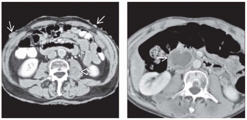
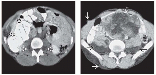
Paraspinal and presacral masses
Plexiform neurofibromas of lumbosacral plexus
Homogeneous, hypodense, cylindrical masses
Often affect iliopsoas compartment
Often involve multiple vertebral levels
Widening of neural foramina
Malignant peripheral nerve sheath tumor
May develop from any plexiform neurofibroma
Most common malignant abdominal tumor in patients with NF1
Large, heterogeneous, variably enhancing mass with ill-defined borders
Plexiform neurofibromas (NF) of: GI, GU tracts; mesentery; hepatobiliary
Other tumors
Ganglioneuroma
Carcinoid tumor (usually duodenal)
Pheochromocytoma and paraganglioma (occur in 1-5%)
GI stromal tumors
Pathology
Schwann cells, fibroblasts, and collagen within myxoid matrix
Accounts for imaging appearance of hypodense, hypovascular masses
Diagnostic Checklist
Most frequent abdominal neoplasm in NF1 is plexiform neurofibroma arising in retroperitoneum, in paraspinal or presacral location
TERMINOLOGY
Abbreviations
Neurofibromatosis type 1 (NF1)
Synonyms
Peripheral neurofibromatosis, von Recklinghausen syndrome (disease)
Definitions
Genetic disorder characterized by development of multiple tumors, predominantly of neuroectodermal or mesenchymal origin
IMAGING
General Features
Best diagnostic clue
Presence of multiple cutaneous and plexiform neurofibromas
Location
Almost anywhere in body
NF1 belongs to group of disorders referred to as “phakomatoses” (neurocutaneous syndromes)
Inherited disorders characterized by tumors or tumor-like nodules of central nervous system, eyes, and skin
Others include
Stay updated, free articles. Join our Telegram channel

Full access? Get Clinical Tree


