
New Directions in Atlas-Assisted Stereotactic Functional Neurosurgery
The first stereotactic brain atlases in printed form, such as Talairach et al and Schaltenbrand and Bailey,1,2 were constructed in the 1950s. Roughly two decades later, brain atlases in electronic formats were available in the clinical setting.3 By the late 1990s, electronic brain atlases had become commonplace in stereotactic functional neurosurgery. The first author and his team have developed the Cerefy electronic brain atlas database, which has become the standard in stereotactic functional neurosurgery. Image-guided surgery companies including Medtronic/Sofamor-Danek, BrainLAB, Cedara/SNN, Elekta, and Integrated Surgical Systems have adopted the Cerefy atlas.
Beginning in the late 1990s, a new-generation brain atlas, referred to as a probabilistic functional atlas (PFA), has been under construction, and a novel way of using it has been proposed. The new atlas is built from electrophysiological and clinical brain mapping data acquired intraoperatively during the treatment of Parkinson’s disease patients. This atlas will be used and its content expanded by the neurosurgical community via an Internet portal, which represents a paradigm shift from a manufacturer-centric to a community-centric atlas. The atlas will become a tool allowing intraoperative targeting based on the patient’s internal landmarks and sufficiently precise to warrant its use for therapeutic purposes. The portal will facilitate data loading, parameter setting, PFA generation and display, and the combination of PFAs and data sets. The atlas and portal are described in greater detail in the section titled Internet Portal for Stereotactic Functional Neurosurgery.
 Electronic Brain Atlas Database
The core of any atlas-assisted application is the brain atlas. Its construction may vary from a simple digitization of a printed atlas to a fully segmented, labeled, enhanced, extended, three-dimensionally expanded, and deformable atlas. We used the latter approach when developing our Cerefy electronic brain atlas database.4,5 This database was derived from the brain atlases edited by Thieme Medical Publishers:
• Atlas for Stereotaxy of the Human Brain (Schaltenbrand and Wahren, 1977)6
• Co-Planar Stereotactic Atlas of the Human Brain (Talairach and Tournoux, 1988)7
• Referentially Oriented Cerebral MRI Anatomy: Atlas of Stereotaxic Anatomical Correlations for Gray and White Matter (Talairach and Tournoux, 1993)8
• Atlas of the Cerebral Sulci (Ono, Kubik, and Abernathey, 1990)9
We digitized these complementary atlases (with gross anatomy, brain connections, subcortical structures, and sulcal patterns) and then enhanced, extended, segmented (color-coded and/or contoured), labeled, aligned, and organized them into atlas volumes. Their three-dimensional extensions were constructed and all two-dimensional (2-D) and three-dimensional (3-D) atlases were mutually co-registered. The combined anatomical index has approximately 1000 structures per hemisphere and more than 400 sulcal patterns.
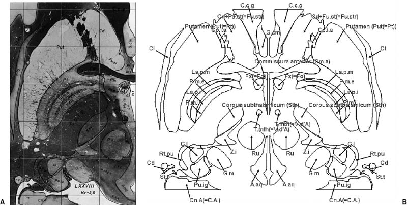
FIGURE 19–1. Schaltenbrand-Wahren brain atlas. (A). Digitized printed axial plate along with the overlay covering the right hemisphere only. (B). Corresponding derived electronic contour image covering both hemispheres with the structures labeled with full or abbreviated names.
The atlases most commonly used in stereotactic functional neurosurgery are the Atlas for Stereotaxy of the Human Brain by Schaltenbrand and Wahren (SW) and the Co-Planar Stereotactic Atlas of the Human Brain by Talairach and Tournoux (TT).
The SW atlas contains photographic plates of macroscopic and microscopic sections through the hemispheres and the brain stem. The microscopic myelin-stained sections show in great detail cerebral deep structures that usually are not well visible on computed tomographic (CT) and magnetic resonance imaging (MRI) scans. The original axial, coronal, and sagittal microseries were digitized with high resolution (Fig. 19–1A). The electronic contours were derived manually from the digitized atlas and labeled. The microseries images and contours were extended to cover both hemispheres. Three SW atlas volumes were constructed with about 600 segmented and labeled structures: coronal with 20 images, sagittal with 34 images, and axial with 20 images (Fig. 19–1B).
The TT atlas was constructed from a single, normal brain specimen (Fig. 19–2A). It had been sectioned and photographed sagittally, and the coronal and axial sections were subsequently interpolated manually. The printed plates were digitized with high resolution, and extensively processed, enhanced, and extended (Fig. 19–2B). The electronic TT atlas images were organized into five atlas volumes: sagittal with 35 images; coronal with 38 images; and three axial with 27 images each, first with standard images, second comprising parcellated cortex (as opposed to the annotated cortex in the original printed atlas) with color-coded Brodmann’s areas on the left side and gyri on the right side, and third containing color-coded gyri on the left side and Brodmann’s areas on the right side. In addition, a contour version of the TT atlas was constructed (Fig. 19–2C).
The 3-D versions of the SW and TT atlases were constructed and mutually co-registered, which enhances surgery planning by providing better insight into spatial relationships (Fig. 19–3).
The electronic atlas images were additionally prelabeled to speed up structure labeling in atlas-based applications. In total, about 17,000 labels were placed manually for the entire electronic brain atlas database (see Figs. 19–1B and 19–2B). Atlas pre-labeling has been used in The Electronic Clinical Brain Atlas10 and Brain Atlas for Functional Imaging.11
 Atlas-Assisted Applications
Electronic brain atlases are commonplace in stereotactic functional neurosurgery. An atlas-assisted application may range from simple (The Electronic Clinical Brain Atlas10) to sophisticated (NeuroPlanner).5,16 Brain atlases are used in various ways, based on several factors:
• Atlas type (2-D SW, 2-D TT, 3-D SW, 3-D TT, others)
• Construction of the computerized atlas (direct digitization of printed plates versus an enhanced, extended, 3-D expanded, and deformable atlas)
• Atlas representation (image, contour, polygonal, volumetric)
• Availability and use of multiple atlases (single atlas, multiple independent atlases, multiple mutually coregistered atlases)
• Atlas-to-data registration method (fully automatic or based on user-specified features such as landmarks or scaling factors)
• Atlas-to-data warping transformation [linear scaling, 3-D piecewise linear scaling (Talairach transformation7) nonlinear warping]
• Availability of interactive atlas-to-data warping (nonavailable; available before planning; available at any time preoperatively, intraoperatively, and postoperatively)
• Atlas display (atlas alone, atlas next to data, atlas images overlaid on data, atlas images overlaid on data with usercontrolled blending, atlas contours overlaid on data)
• Atlas-assisted operations (atlas labeling, data labeling, multiple orientation targeting, multiple atlas targeting, planning in 2-D and 3-D on the atlas-segmented anatomy)
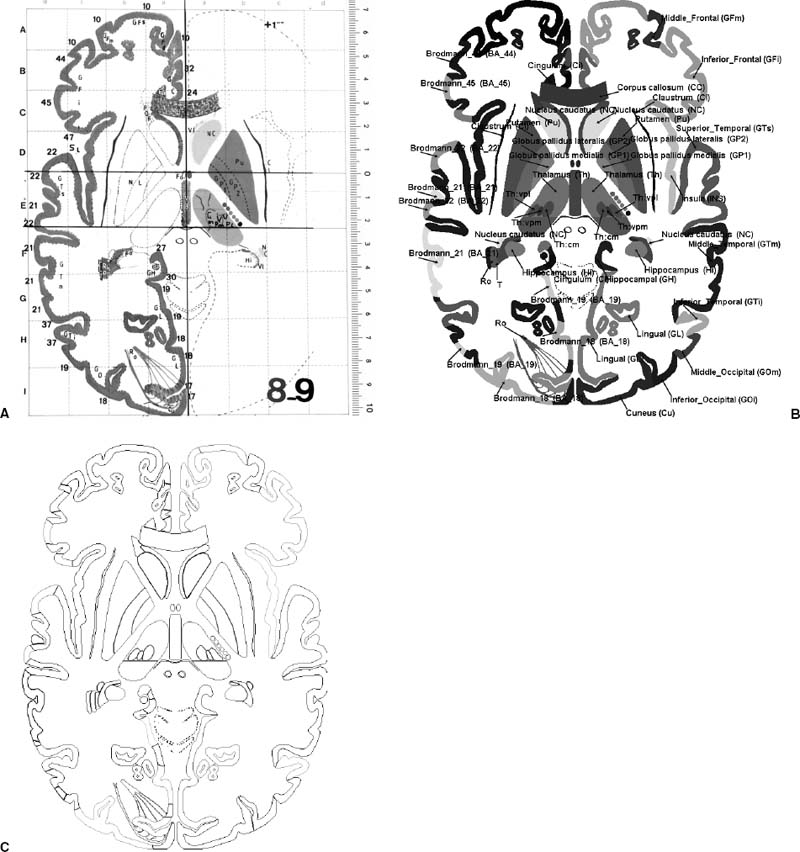
FIGURE 19–2. Talairach-Tournoux brain atlas. (A). Digitized printed axial plate; (B) corresponding electronic color-coded image labeled with subcortical structures, gyri, and Brodmann’s areas (full or abbreviated names are used); (C) corresponding color-coded contours.
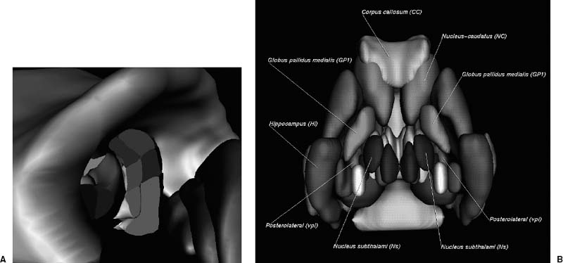
FIGURE 19–3. Three-dimensional atlases. (A). Talairach-Tournoux (TT) atlas co-registered with the Schaltenbrand-Wahren (SW) atlas: a view into the thalamic nuclei of the 3-D SW atlas combined with the basal ganglia and corpus callosum of the 3-D TT atlas. (B). Subcortical structures of the 3-D Talairach-Tournoux atlas with the stereotactic targets labeled on both sides.
Cerefy brain atlas applications
The Electronic Clinical Brain Atlas (ECBA)10 on CD-ROM contains, among others, the SW and TT atlases. The ECBA provides features not available in the printed atlases such as: mutually co-registered atlases; fully pre-labeled 17,000 structures on 1500 atlas images; flexible display, manipulation, and printing of the atlas in multi-atlas and triplanar modes; and atlas warping. The ECBA generates individualized atlases without loading the patient-specific data, which is useful for targeting. The ECBA-based planning procedure for stereotactic functional neurosurgery has been described.13 The atlas is conformed to the patient’s scan by means of a 2-D local deformation done by matching the atlas rectangular region of interest to the corresponding data region of interest spanned on any landmarks. The deformation can be repeated in multiple orientations, if data are available, increasing the accuracy of targeting and the neurosurgeon’s confidence level.
Two add-on brain atlas libraries are gaining increasing acceptance and use, the Electronic Brain Atlas Library and Brain Atlas Geometrical Models. The Electronic Brain Atlas Library (EBAL)14 contains the brain atlas database with the SW and TT atlas images, and a browser. The browser provides means for exploring and understanding the atlas images and allows users to build their own atlasassisted applications. It also allows the user to select atlas volume, display atlas images, find labels of structures, display atlas image location in 3-D space, display stereotactic grids, find stereotactic coordinates, and search for structure. These features also make the EBAL useful as a stand-alone atlas reference.
The Brain Atlas Geometrical Models15 is a library with the atlases in contour and polygonal representations. The BAGM contains the brain atlas database and a viewer. The database comprises the SW atlas in contour representation and 3-D polygonal models of the SW and TT atlases. The viewer provides means for viewing and understanding the brain atlas database.
The detailed specifications of the EBAL content and the BAGM content along with file formats are available online at www.cerefy.com.
The NeuroPlanner5,16 is a clinical prototype developed for preoperative planning and training, intraoperative procedures, and postoperative follow-up. It comprises all (mutually co-registered) atlases from the Cerefy brain atlas database, including their 3-D extensions. The atlases are available in image, contour, and polygonal representations as well as in multiple resolutions. The atlas-to-data registration is based on the Talairach landmarks specified in the data interactively; alternatively, the placement of the landmarks can be done automatically.12 The NeuroPlanner
Related posts:
 Intraoperative Image Update by Interface with Ultrasound
Stay updated, free articles. Join our Telegram channel

Full access? Get Clinical Tree


