(1)
Kaiser Permanente, Southern California Permanente Medical Group, Riverside, CA, USA
(2)
Molecular Imaging Center and PET Clinic, University of Southern California, Los Angeles, CA, USA
(3)
Keck School of Medicine, University of Southern California, Los Angeles, CA, USA
18F-AV-45 and 18F-FDG
Case 17.1
Findings
Case 17.2
Findings
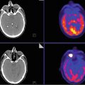
The 18F-FDG brain PET images of the brain (Fig. 17.2, top) demonstrate bilateral temporal hypometabolism consistent with dementia of Alzheimer’s type by pattern and the 18F-AV-45 PET images of brain (Fig. 17.2, bottom) demonstrate diffuse uptake in the brain with loss of gray and white matter demarcation unlike in case 1 (Fig. 17.2).


Fig. 17.2

